HOME > Weekly Case > Weekly Case
Weekly Case
종결
Case No. 097
0000-00-00~0000-00-00
출제자 : 관리자
Hit : 336
| Period | 0000-00-00~0000-00-00 |
|---|---|
| Diagnosis | |
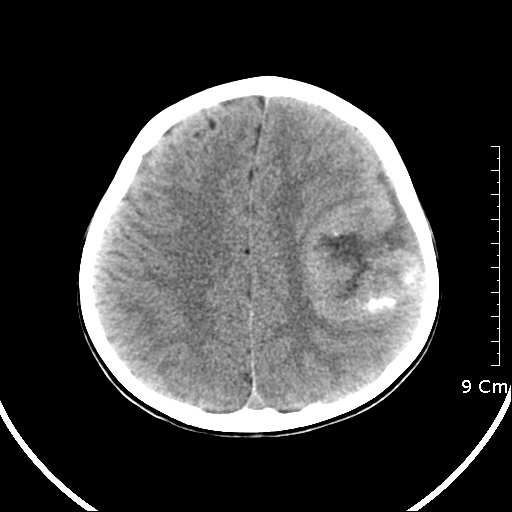
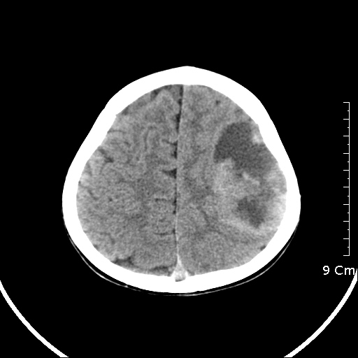
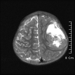
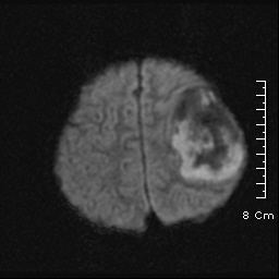
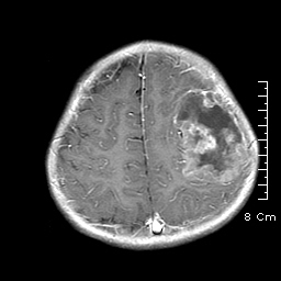
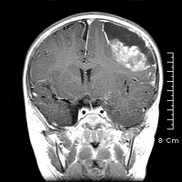
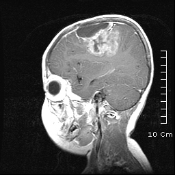
| Clinical information | 3세 여아, Seizure로 내원. |
| Discussion | [정답] Supratentorial ependymoma 1) 40 % of ependymoma, older age than infratentorial group (18 – 24 yrs) 2) different clinical presentation: supratentorial vs infratentorial ependymoma -> Supratentorial ependymomas: focal neurologic deficits, headache, & seizures -> Infratentorial ependymoma: intraventricular location, clinically manifest earlier (increased intracranial pressure & hydrocephalus) 3) Imaging (1) Supratentorial ependymoma: -> brain parenchyma (embryonic rests of ependymal tissue trapped in the developing cerebral hemispheres) -> Owing to its parenchymal location, larger in size than its infratentorial counterpart (>4 cm) -> cystic component (2) Infratentorial ependymoma: -> Intraventricular, smaller, more often solid (3) Both Supratentorial & Infratentorial -> Calcification: very common in both infra- & supratentorial ependymomas (40%–80% of cases) -> Same imaging characteristics at CT and MR imaging: - iso- to slightly hypoattenuating at unenhanced CT - iso-to hypointense on unenhanced T1-weighted MR images and hyper-intense on T2- & proton-density–weighted MR images - variable contrast enhancement behavior with central areas of necrosis (4) D/Dx with the appearance of an extraventricular supratentorial ependymoma -> astrocytoma (both low grade and glioblastoma multiforme), supratentorial primitive neuroectodermal tumor, gangliogliomacytoma, & oligodendroglioma |
| Correct answer |
Correct AnswerSemi-Correct Answer |