HOME > Weekly Case > Weekly Case
Weekly Case
종결
Case No. 093
0000-00-00~0000-00-00
출제자 : 관리자
Hit : 384
| Period | 0000-00-00~0000-00-00 |
|---|---|
| Diagnosis | |
| Clinical information | Facial numbness로 내원한 59세 남자. |
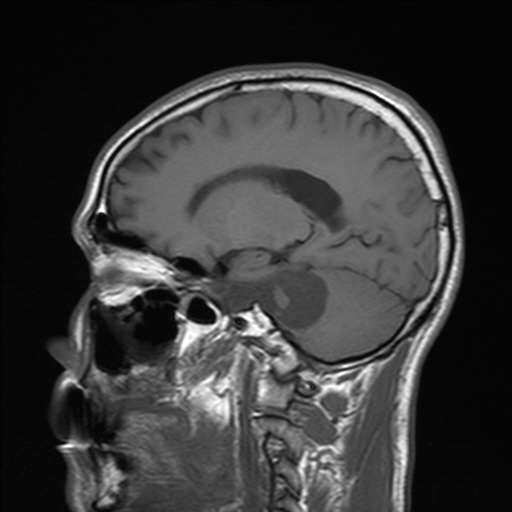
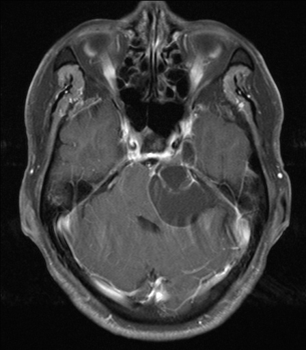
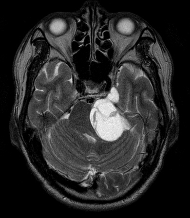
| Discussion | [정답] Trigeminal Schwannoma Image findings: 2005-6-16 MR shows a 3.5 cm sized multiseptated cystic mass with extension to Meckel's cave in the left CPA. The mass extends to pons superiorly, and medulla inferiorly. Brain stem shift to right side by the mass. Pathologic finding: Pathologic report said that the pathologic specimen were grossly multiple fragments of pinkish brown soft tissue, measuring up to 1.5cm in greatest dimension. and they were consistent with schwannoma. Discussion Arise from CPA, Meckel's cave, gasserian ganglion in the middle cranial fossa. Involve both the posterior and middle cranial fossa. Centered anteromedial to the IAC, extends into Meckel's cave. Foramen ovale or the foramen rotundum also may be enlarged. More likely to contain cystic components than acoustic schwannoma. CT findings: iso to cerebellum, calcification or hemorrhge is rare. Homogeneous and dense enhancement. Some show irregular central lucencies or inhomogeneous enhancement (cystic component). MR imaging : T1WI isointense or mildly hypointense relative to the pons/ hyperintense to CSF. T2WI mildly hyperintense to the pons/ iso to hypointense to CSF. Intense enhancement. References: PeterM. Som Hugh D. Curtin et al. Head and neck imaging 4th ed. Mosby 2003;1291-1293 |
| Correct answer |
Correct AnswerSemi-Correct Answer |