HOME > Weekly Case > Weekly Case
Weekly Case
종결
Case No. 092
0000-00-00~0000-00-00
출제자 : 관리자
Hit : 416
| Period | 0000-00-00~0000-00-00 |
|---|---|
| Diagnosis | |
| Clinical information | 63세 남자. 2개월간의 gait disturbance, headache, memory dysfunction으로 내원. |
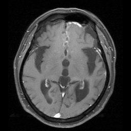
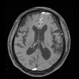
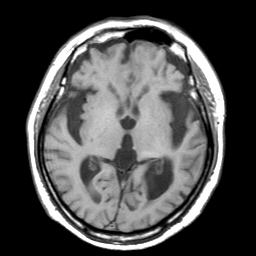
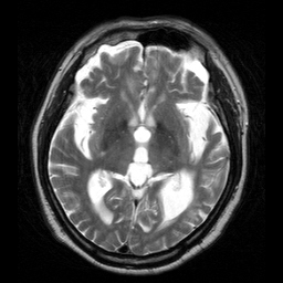
| Discussion | [Answer] Neurocysticercosis 1. Widening of both sylvian fissure, focal cystic lesion at left frontal pole area. 2. Meningitis along anterior part of interhemispheric fissure. 3. Enhancing degenerative calcified granuloma at the left frontal and left parietal lobe. 4. Communicating hydrocephalus. 1. Etiology The larval form of the pork intestinal parasite:Taniae solium is the agent responsible for Neurocysticercosis. 2. Incidence 1) Neurocysticercosis is the most common CNS parasitic infection. 2) CNS involvement occurs in 60-90% of patients with cysticercosis. 3) It is endemic in many areas such as Central and South America, Eastern Europe, Africa, and parts of Asia. 3. Location 1) The brain parenchyma is the most commonly affected site in NCC and the corticomedullary junction is the primary location. 2) Intraventricular cysticercosis cysts are seen in 20-50% of case with the fourth ventricle a common site. 3)Only 10 % of NCC cases have isolated subarachnoid disease. 4. Clinical presentaion and natural history NCC has a broad spectrum of clinical manifestations. Epilepsy is the most frequent symptom and is seen in 50-70%. 5. Pathology and imaging correlation The pathologic manifestations of parenchymal cysticercosis have been classified into the following four stages: 1) Vesicular stage : thin capsule, fluid containing. Edema and contrast enhancement are rare. 2) Colloid vesicular state : larvum dies and begins to degeneration, edema and cystic wall enhancement. 3) Granular nodular state : capsule thickening, calcification 4) Nodular calcification state : small calcificed nodule without mass effect or enhancement. 6.Treatment: Praziquantel |
| Correct answer |
Correct AnswerSemi-Correct Answer |