HOME > Weekly Case > Weekly Case
Weekly Case
종결
Case No. 759
2026-08-01~2026-08-31
출제자 : 전영훈
Hit : 206
| Period | 2026-08-01~2026-08-31 |
|---|---|
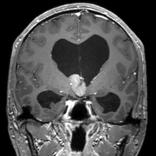
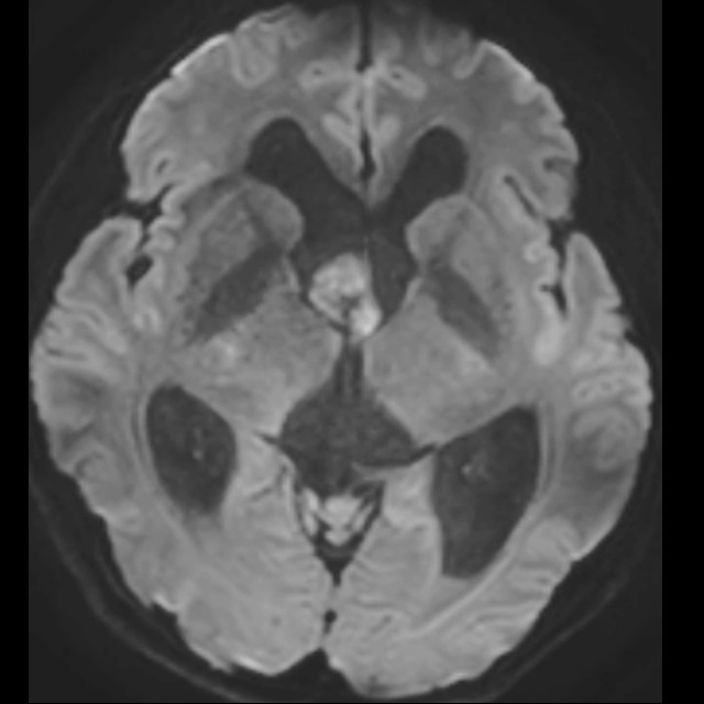
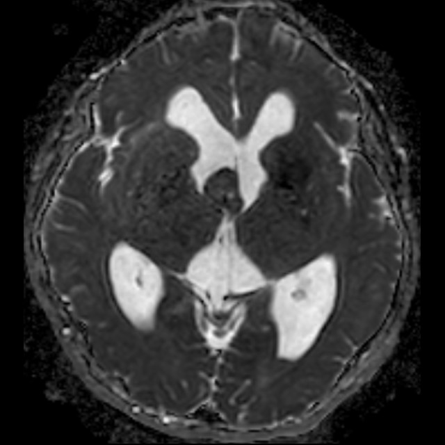
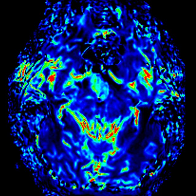
| Diagnosis | Intraventricular meningioma |
| Gender |
|
| Age | 34 |
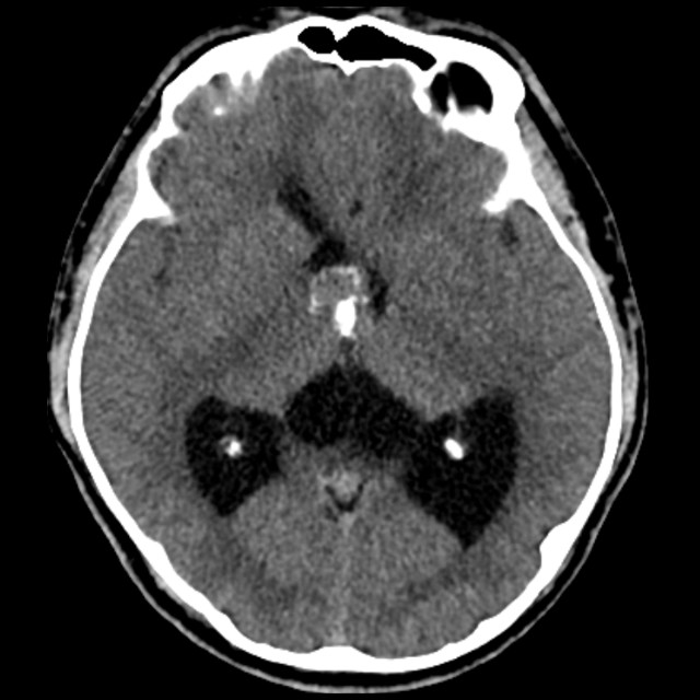
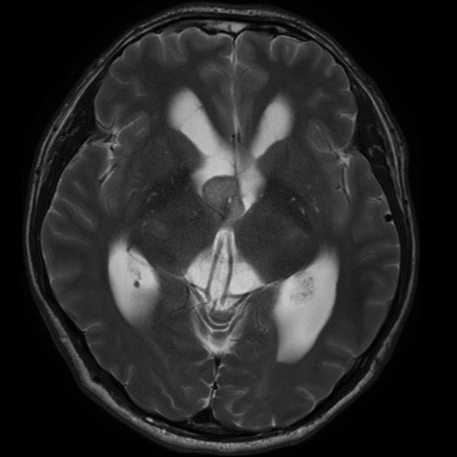
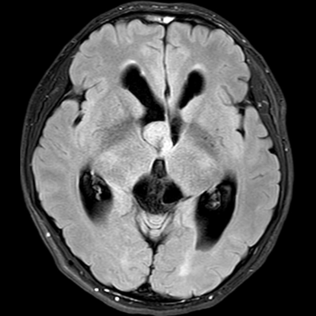
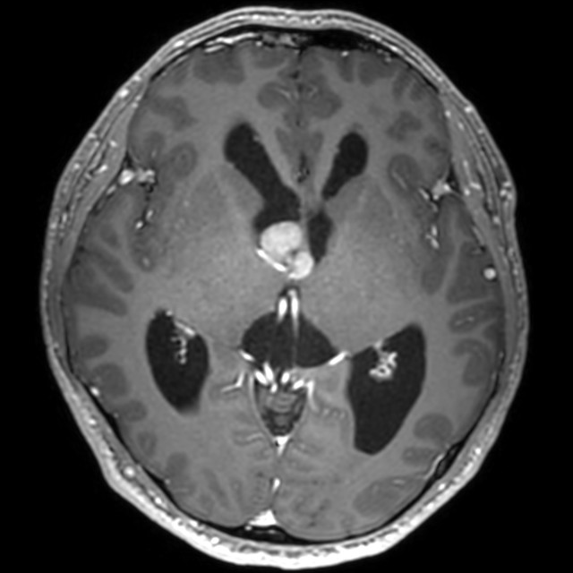
| Clinical information | Brain MRI of a 34-year-old male who presented with syncope. What is your diagnosis? |
| Discussion | Epidemiology and Location approximately 1–5% of all meningiomas 30–60 years, female predominance Approximately 90% are WHO grade 1 Most commonly located in the lateral ventricular atrium (trigone) Imaging Features Well-circumscribed, lobulated intraventricular mass Usually iso- to hypointense on T1- and T2-weighted images Strong, homogeneous contrast enhancement Calcification may be present on CT Typically hypervascular, supplied mainly by the anterior and/or posterior choroidal arteries DDx.: Central neurocytoma Septum pellucidum near the foramen of Monro Heterogeneous, bubbly appearance with cystic change and calcification Mild to moderate and heterogeneous enhancement Reference Niiro T, Tokimura H, Hanaya R, et al. MRI findings in patients with central neurocytomas with special reference to differential diagnosis from other ventricular tumours near the foramen of Monro. J Clin Neurosci. 2012 May;19(5):681-6. |
| Correct answer |
Correct Answer
Semi-Correct Answer |
Submit answer
Answer submission list
The written answers can only be viewed by the administrator and author.