HOME > Weekly Case > Weekly Case
Weekly Case
종결
Case No. 749
2026-03-01~2026-03-31
출제자 : 신일아
Hit : 578
| Period | 2026-03-01~2026-03-31 |
|---|---|
| Diagnosis | Middle ear neuroendocrine tumor (middle ear adenoma) |
| Gender |
|
| Age | 35 |
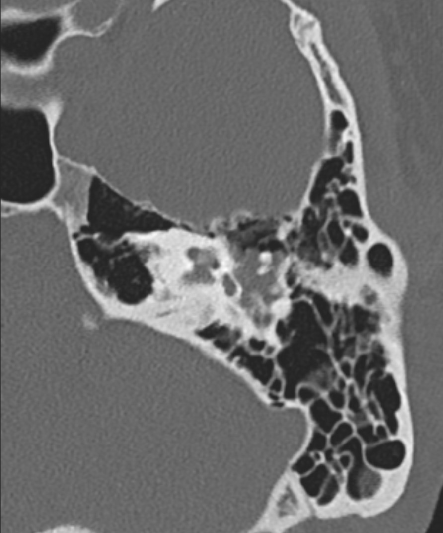
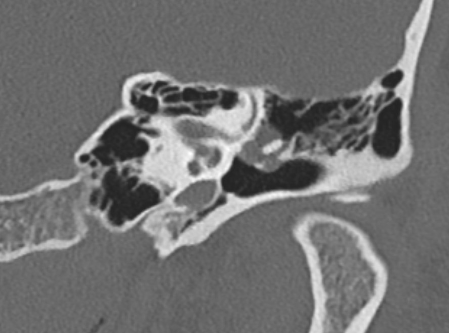
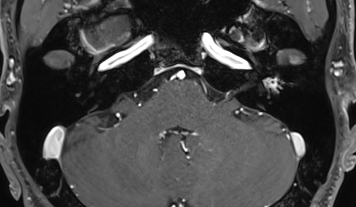
| Clinical information | M/35 Chief complaint) Ear fullness, left for 3 month Past history) 어렸을 적 COM history 있음 A 35-year-old male patient with no significant past medical history other than a history of otitis media in childhood, presenting with a chief complaint of left ear fullness persisting for the past three months. What is the diagnosis? |
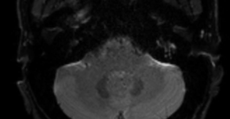
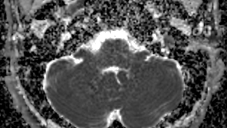
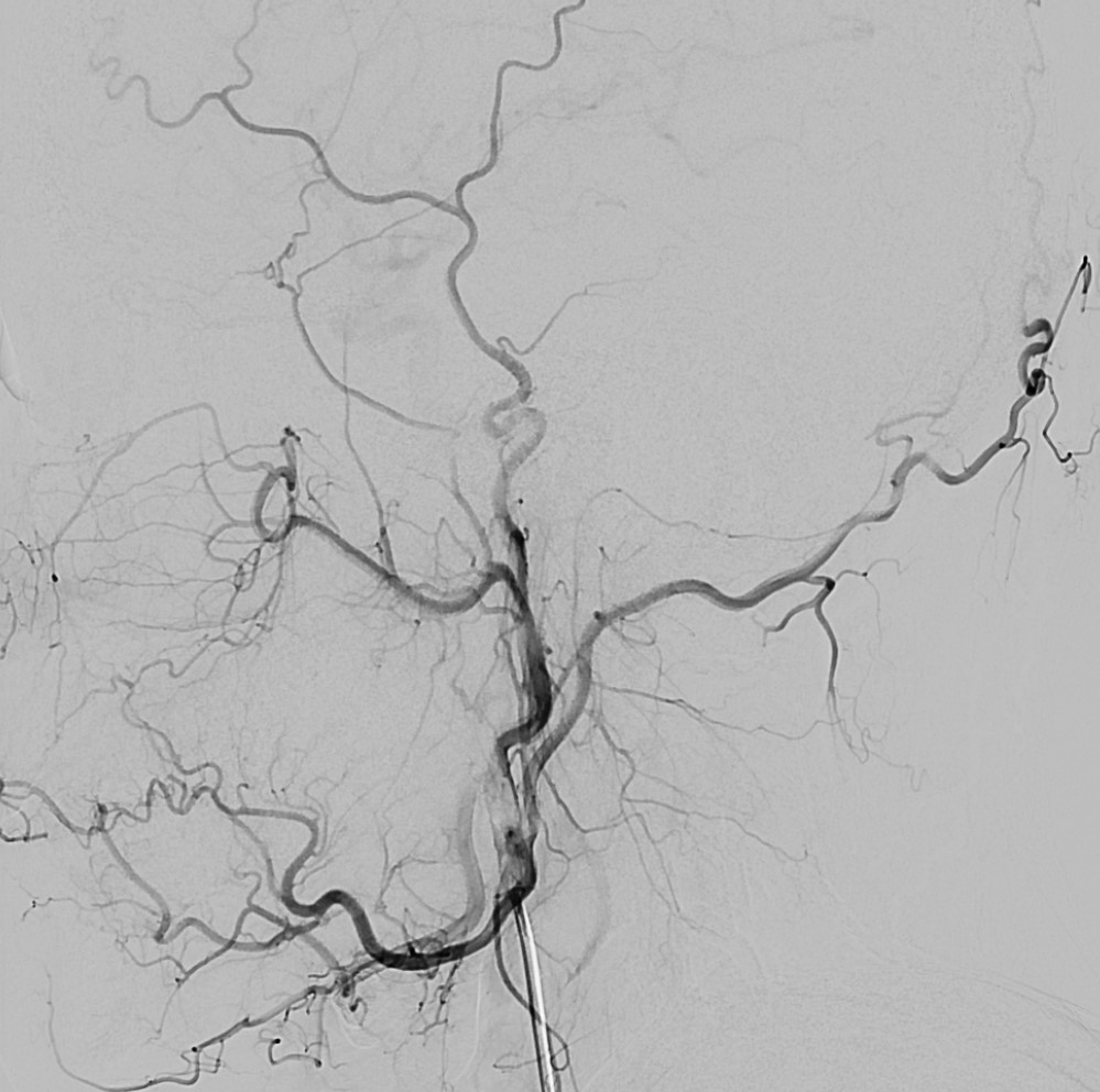
| Discussion | Imaging demonstrates a well-defined soft tissue mass within the middle ear cavity that surrounds the ossicular chain, particularly the malleus–incus complex, without evidence of ossicular erosion. On MRI, the lesion shows relatively homogeneous enhancement and mild diffusion restriction in this case, while digital subtraction angiography reveals no hypervascular tumor blush. These imaging findings favor Middle ear adenoma, a rare benign neoplasm arising from the middle ear mucosa. Middle ear adenomas typically present as non-destructive soft tissue masses that may envelop the ossicles while preserving the ossicular structures, often manifesting clinically with symptoms such as ear fullness, tinnitus, or conductive hearing loss. An important differential consideration is Glomus tympanicum, the most common primary neoplasm of the middle ear. In contrast to middle ear adenoma, glomus tympanicum is a highly vascular tumor that typically arises from the cochlear promontory along the tympanic branch of the glossopharyngeal nerve (Jacobson nerve). Imaging usually demonstrates avid enhancement and a prominent tumor blush on angiography, and larger lesions may exhibit internal flow voids producing a “salt-and-pepper” appearance on MRI. Moreover, glomus tympanicum lesions are usually promontory-centered, whereas middle ear adenomas more commonly encase the ossicular chain without a clear promontory origin. In the present case, presence of a non-erosive ossicle-encasing mass centered around the malleus–incus complex and negative DSA strongly favor the diagnosis of middle ear adenoma over glomus tympanicum. |
| Correct answer |
Correct AnswerSemi-Correct Answer |
Submit answer
Answer submission list
The written answers can only be viewed by the administrator and author.
The written answers can only be viewed by the administrator and author.