HOME > Weekly Case > Weekly Case
Weekly Case
종결
Case No. 747
2026-03-01~2026-03-31
출제자 : 신일아
Hit : 415
| Period | 2026-03-01~2026-03-31 |
|---|---|
| Diagnosis | Ganglioglioma |
| Gender |
|
| Age | 18 |
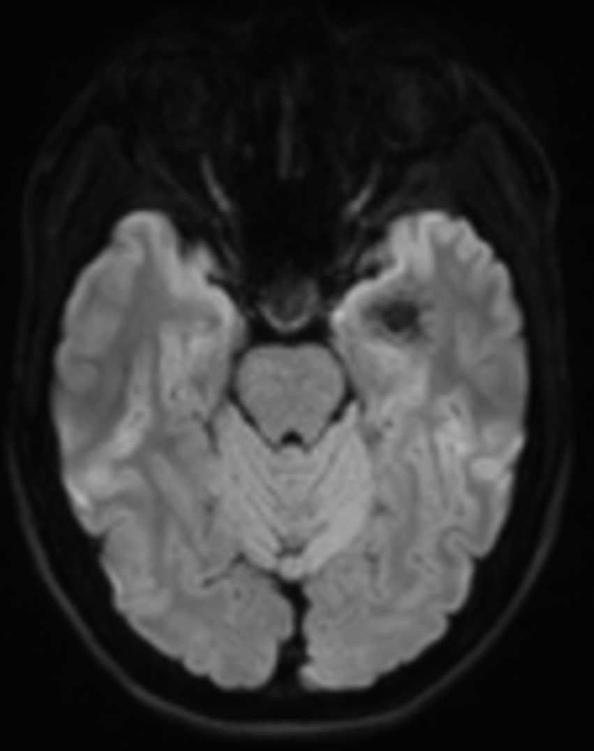
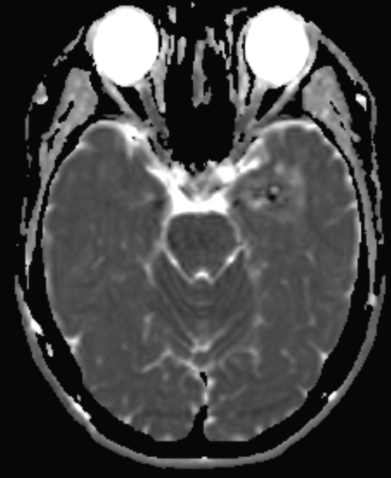
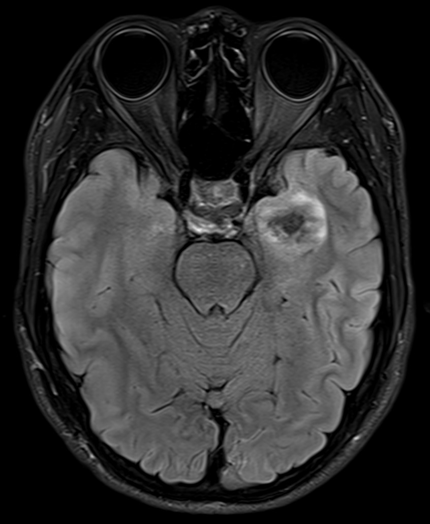
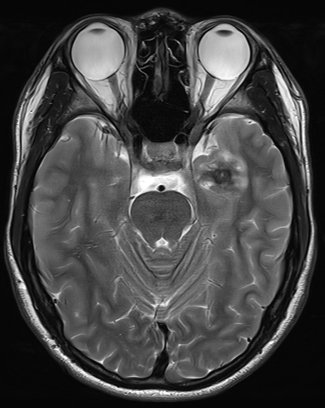
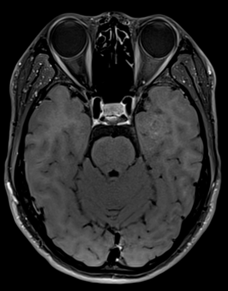
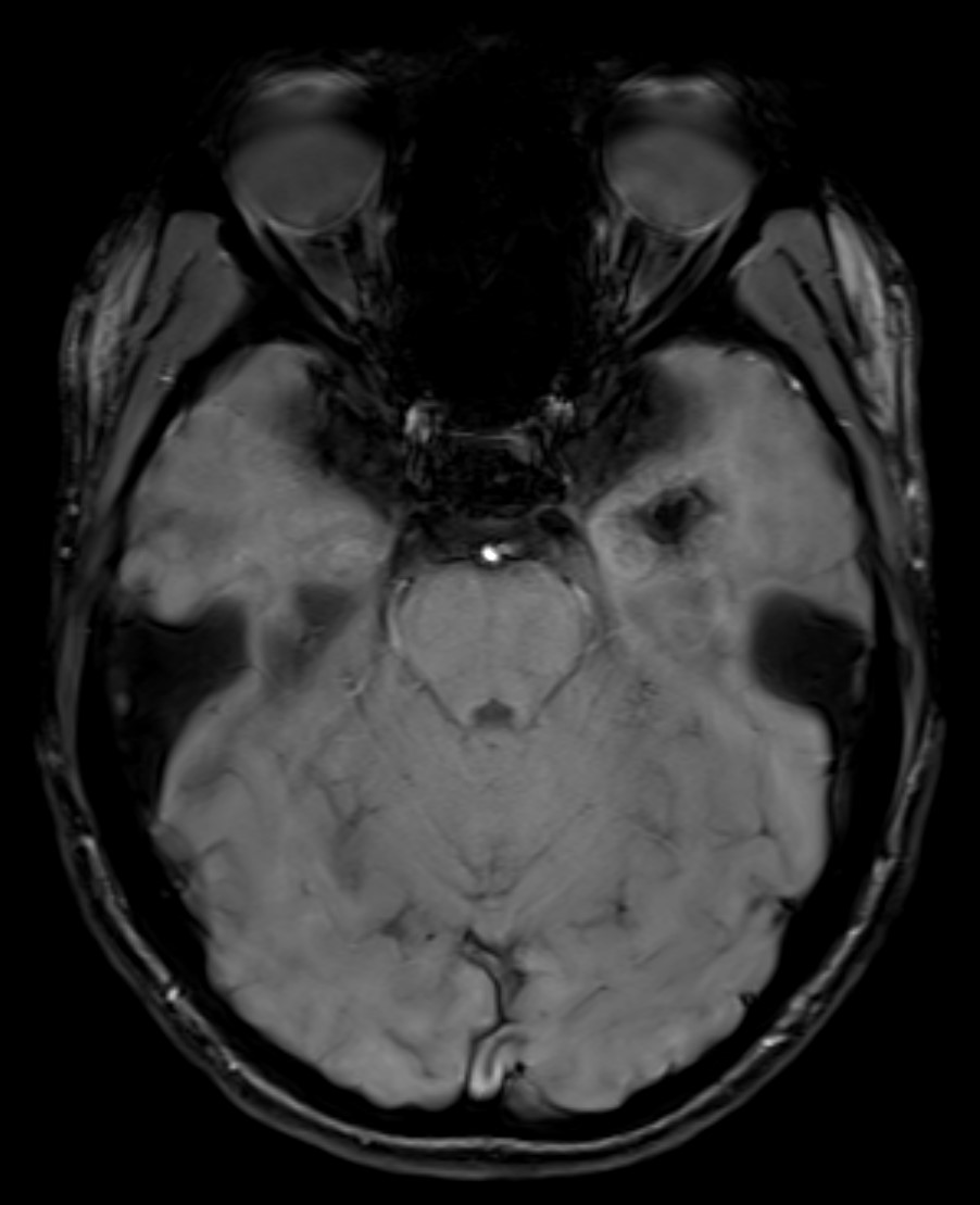
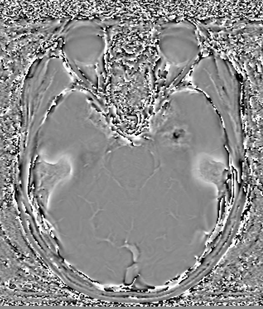
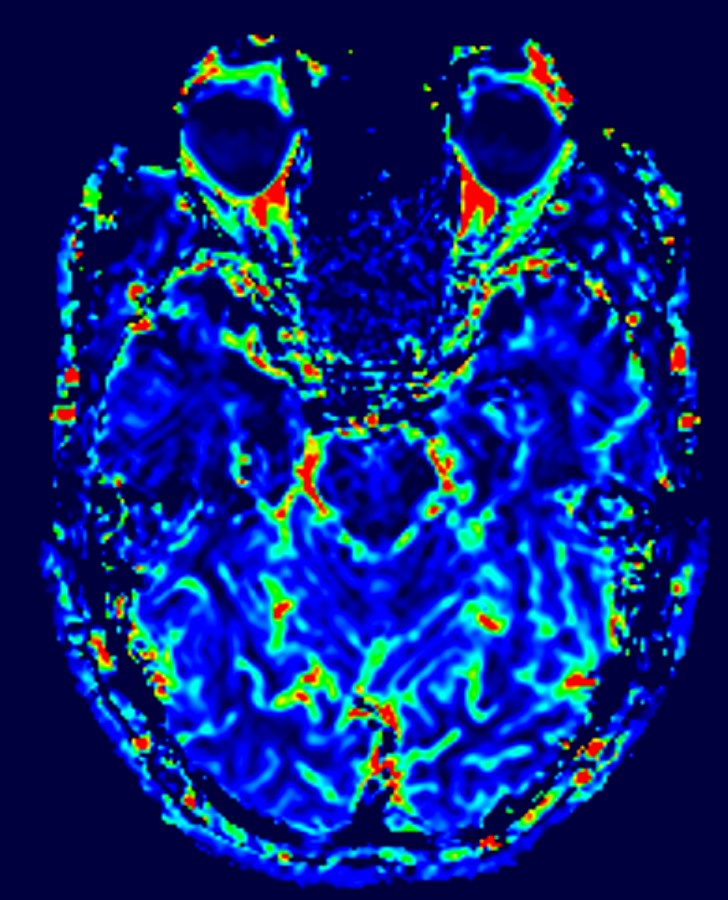
| Clinical information | M/18, Chief complaint) none – incidental finding An 18-year-old male patient with no significant medical history or underlying diseases visited for evaluation of a mass incidentally found during a check-up. What is the diagnosis? |
| Discussion | Ganglioglioma is a rare, typically low-grade glioneuronal tumor composed of neoplastic ganglion cells and neoplastic glial components. It most commonly arises in the temporal lobe and frequently involves the cortical and subcortical regions. These tumors usually occur in children and young adults and are often associated with long-standing epilepsy; however, incidental detection in asymptomatic patients may also occur. Histologically, most gangliogliomas correspond to WHO grade 1, although anaplastic transformation is rare. Ganglioglioma demonstrates a wide spectrum of imaging appearances because of its mixed neuronal and glial cellular composition and variable stromal components. On MRI, gangliogliomas are usually well-circumscribed lesions that show variable signal intensity. On T1-weighted imaging, the lesion is generally iso- to hypointense relative to gray matter, while T2-weighted and FLAIR images typically demonstrate hyperintensity, however, in the present case, the lesion demonstrated low signal intensity on both T1- and T2-weighted images, likely attributable to internal calcifications. Peritumoral edema and mass effect are often minimal relative to tumor size, reflecting the generally indolent growth pattern. Enhancement patterns are highly variable. Some tumors show no enhancement, whereas others demonstrate focal, heterogeneous, or nodular enhancement. Calcification is frequently present and may be detected more clearly on CT (however, CT was not available in our case). Cystic components are also common, sometimes with an enhancing mural nodule. Less commonly, the lesion may appear predominantly solid, partially cystic, or demonstrate mixed signal characteristics due to calcification, cyst formation, or occasional hemorrhage. Because of this variability, the imaging differential diagnosis often includes other cortical-based low-grade tumors such as oligodendroglioma, dysembryoplastic neuroepithelial tumor (DNET), and other pediatric-type low-grade gliomas. |
| Correct answer |
Correct Answer
Semi-Correct Answer |
Submit answer
Answer submission list
The written answers can only be viewed by the administrator and author.