HOME > Weekly Case > Weekly Case
Weekly Case
종결
Case No. 746
2026-02-01~2026-02-28
출제자 : 김정윤
Hit : 605
| Period | 2026-02-01~2026-02-28 |
|---|---|
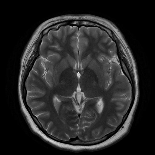
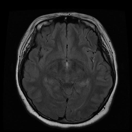
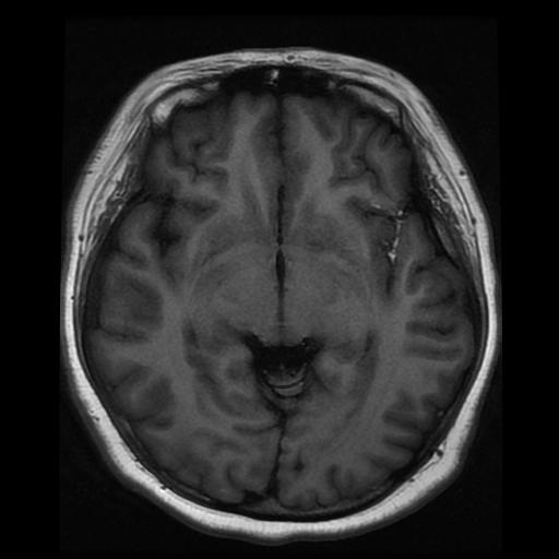
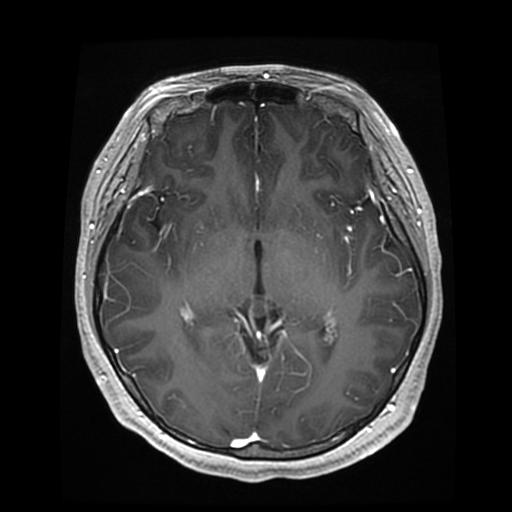
| Diagnosis | Ruptured intracranial dermoid cyst |
| Gender |
|
| Age | 19 |
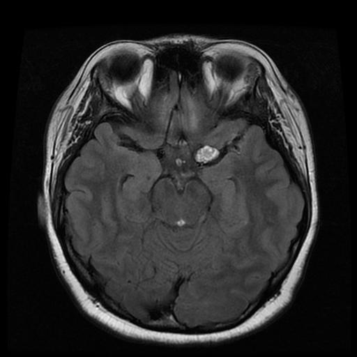
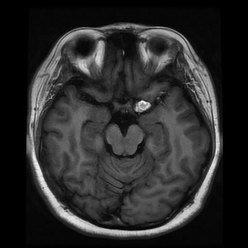
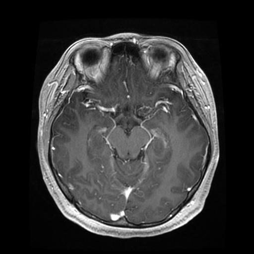
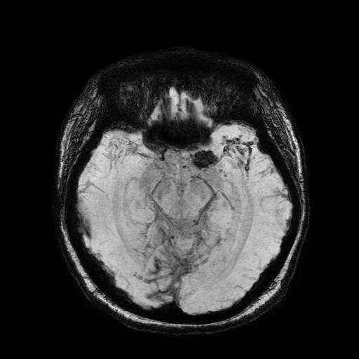
| Clinical information | A 19-year-old female patient presented with a headache. What was the diagnosis? |
| Discussion | A ruptured intracranial dermoid cyst is a rare complication of a congenital intracranial tumor caused by leakage of lipid-rich contents into the subarachnoid space or ventricular system. Clinically, patients most commonly present with headache and seizure, and rupture may lead to chemical meningitis. On MRI, unruptured dermoid cysts typically show marked T1 hyperintensity due to lipid content. After rupture, scattered T1-hyperintense fat droplets are characteristically seen within the subarachnoid spaces and/or ventricles, with confirmation on fat-suppressed sequences. Signal intensity on T2-weighted images is variable, and mild peripheral capsular enhancement may be present. Leptomeningeal enhancement can be seen in ruptured cases, reflecting chemical meningitis. Reference Osborn AG, Preece MT. Intracranial cysts: radiologic-pathologic correlation and imaging approach. Radiology. 2006;239 (3): 650-64. |
| Correct answer |
Correct Answer
Semi-Correct Answer |
Submit answer
Answer submission list
The written answers can only be viewed by the administrator and author.