HOME > Weekly Case > Weekly Case
Weekly Case
종결
Case No. 745
2026-02-01~2026-02-28
출제자 : 김정윤
Hit : 478
| Period | 2026-02-01~2026-02-28 |
|---|---|
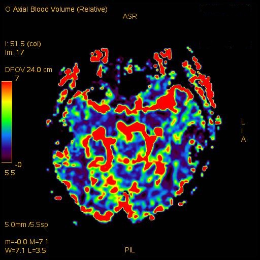
| Diagnosis | Radiation-induced organizing hematoma |
| Gender |
|
| Age | 48 |
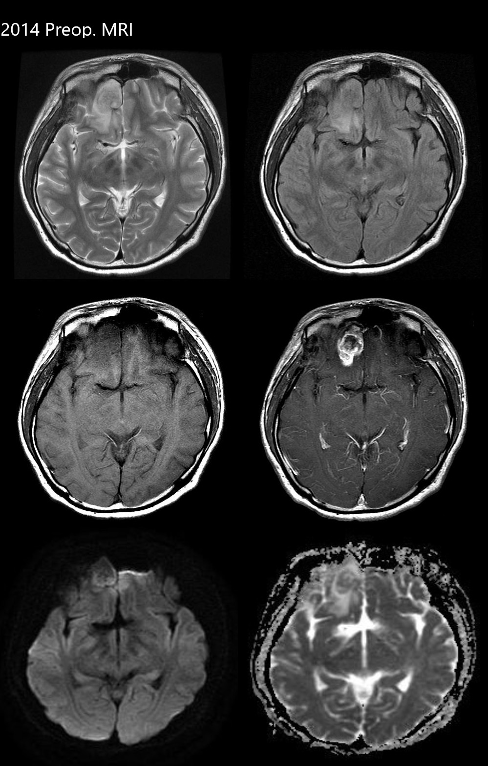
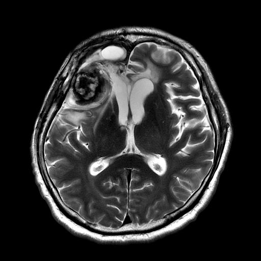
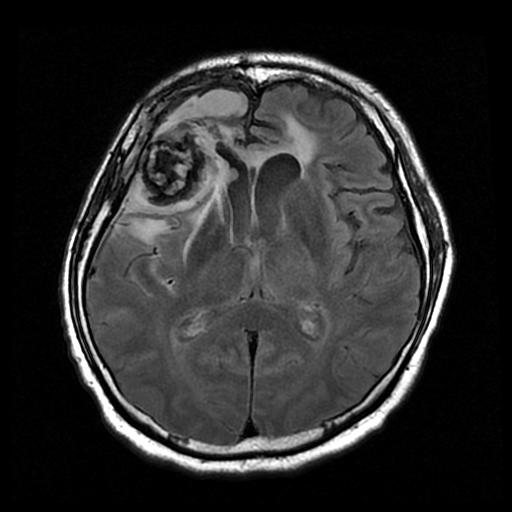
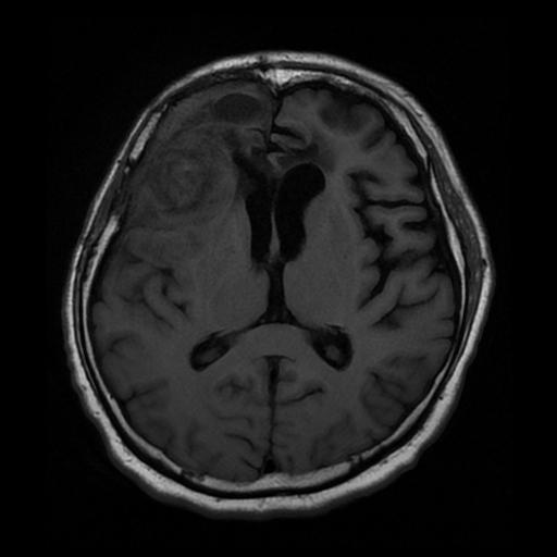
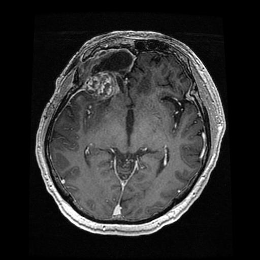
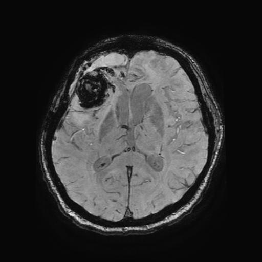
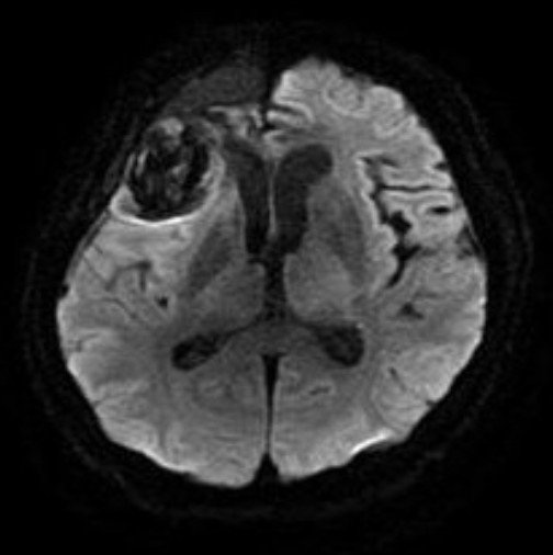
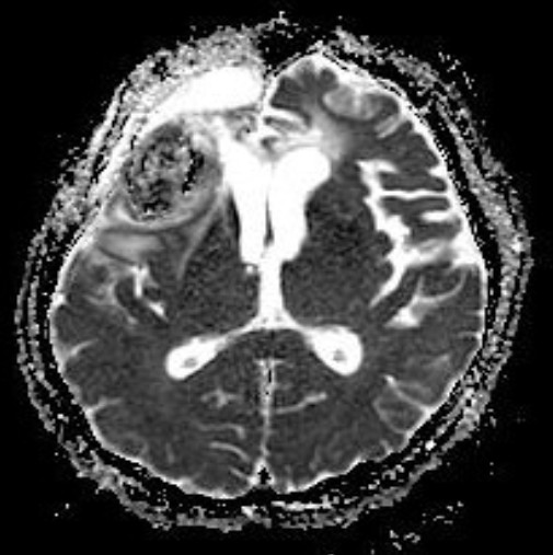
| Clinical information | A 48-year-old male patient presented with nausea after undergoing tumor removal and radiation treatment for a brain mass in the right frontal lobe in November 2014 (approximately 10 years ago). What is his current diagnosis? |
| Discussion | Radiation-indicated organizing hematoma (RIOH) refers to a localized vascular, tumor-like lesion that develops after cerebral radiation. The lesion occurs mainly in children and young people, with variable latency periods after radiation treatment and diverse original lesions, including arteriovenous malformation, glioma, and metastatic tumor. Similar to de novo cavernous hemangioma, RIOH appears as an enhancing lesion with popcorn-like appearance and partial hemosiderin rim on magnetic resonance imaging (MR) and is observed histologically as a vascular-rich hemorrhagic lesion. MR imaging usually shows a reticulated core of heterogeneous signal intensity with a dark peripheral rim of hemosiderin, giving a typical “popcorn” appearance. CT usually shows ring-like calcification with core reticulation of variable attenuation. Reference Kim MS, Kim SH, Chang JH, Park M, Cha YJ. Clinicopathological differences in radiation-induced organizing hematomas of the brain based on type of radiation treatment and primary lesions. J Pathol Transl Med. 2022;56(1):16-21. Jain R, Robertson PL, Gandhi D, Gujar SK, Muraszko KM, Gebarski S. Radiation-induced cavernomas of the brain. AJNR Am J Neuroradiol. 2005 May;26(5):1158-62. |
| Correct answer |
Correct Answer
Semi-Correct Answer |
Submit answer
Answer submission list
The written answers can only be viewed by the administrator and author.