HOME > Weekly Case > Weekly Case
Weekly Case
종결
Case No. 732
2025-10-01~2025-10-31
출제자 : 정소영
Hit : 384
| Period | 2025-10-01~2025-10-31 |
|---|---|
| Diagnosis | Diffuse scalp neurofibroma |
| Gender |
|
| Age | 44 |
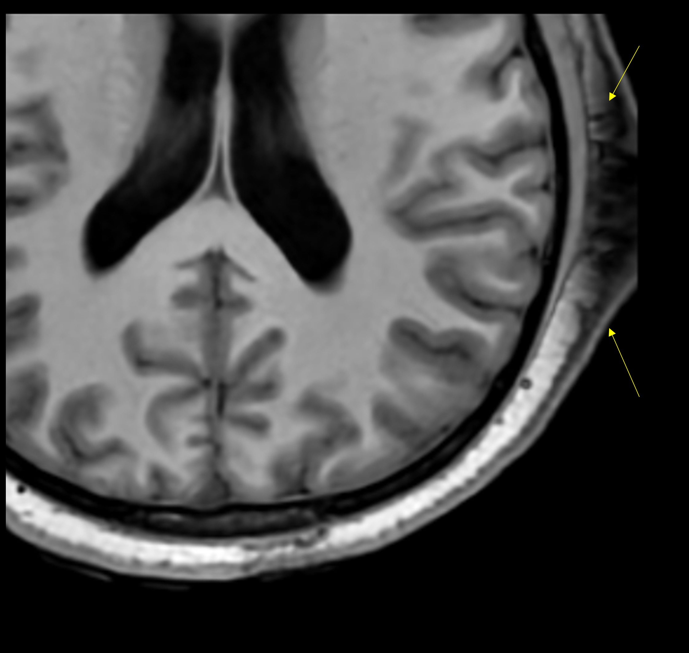
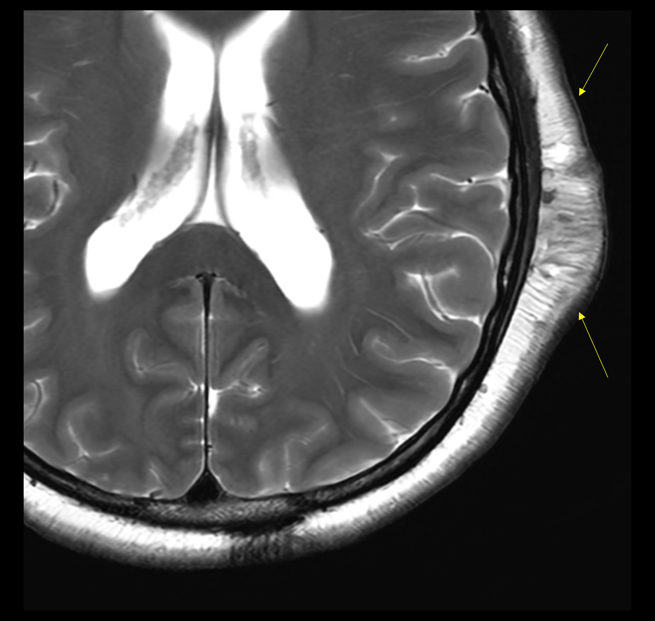
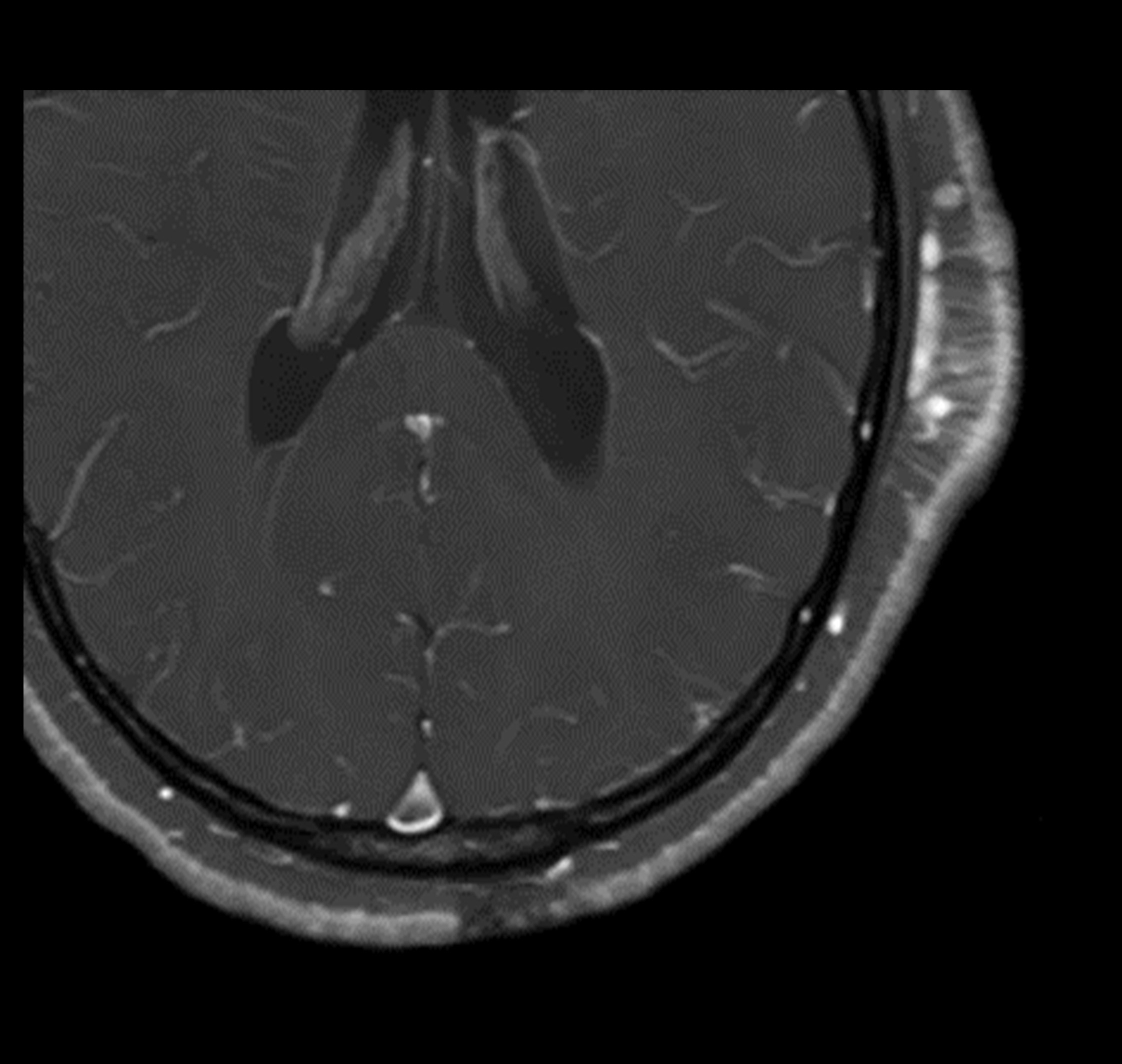
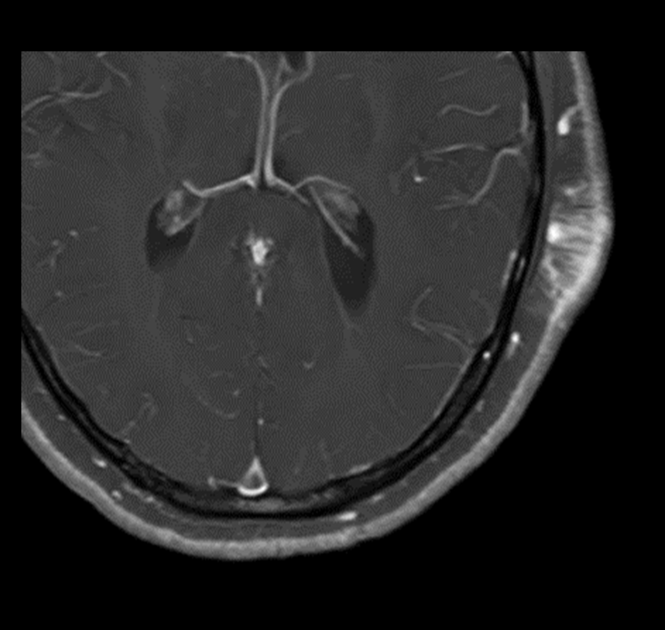
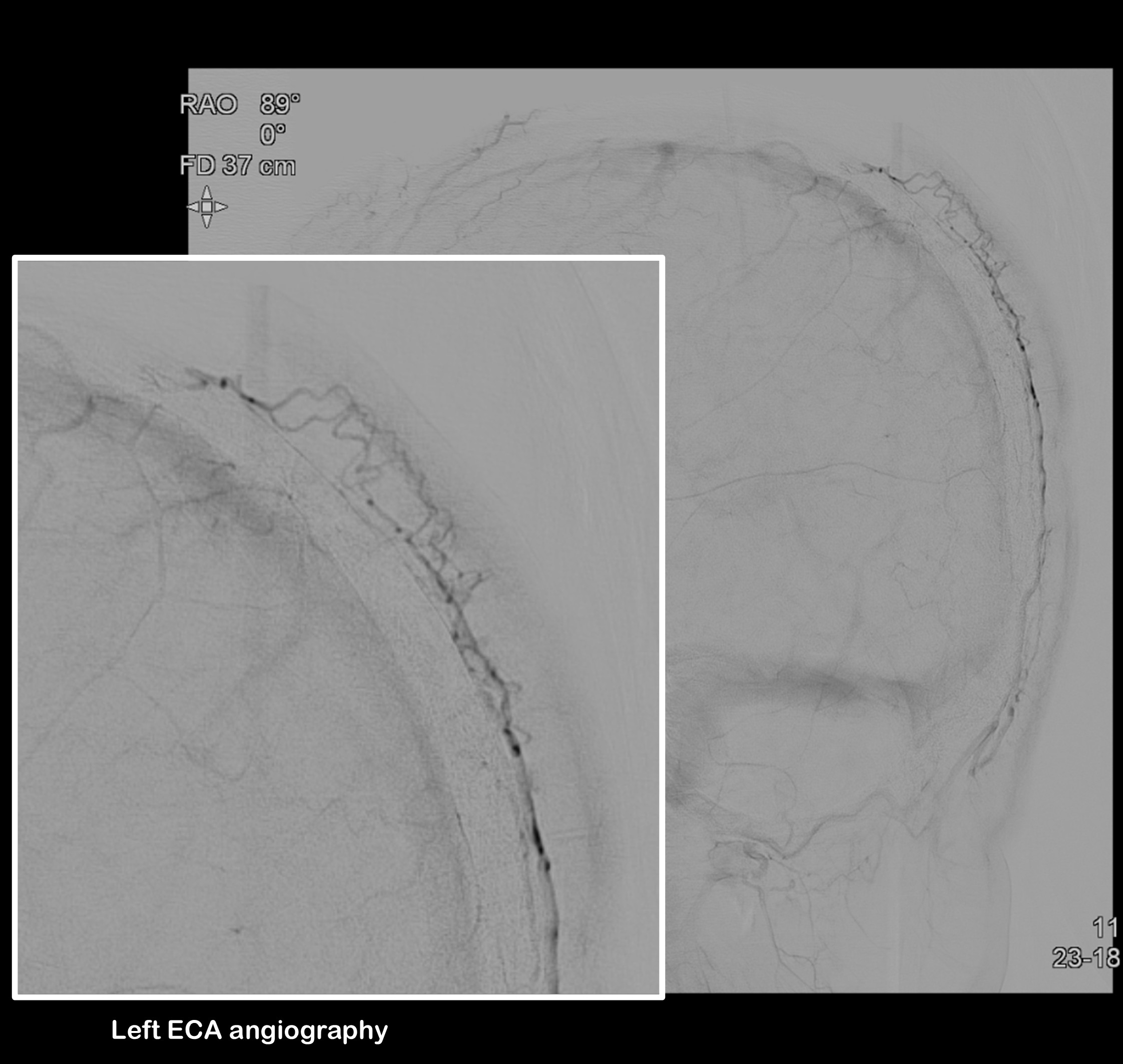
| Clinical information | F/44Y Symptom: Scalp lesion On physical examination, a mass-like lesion was palpated on the left scalp, where there is also a localized patch of white hair consistent with poliosis. Images> T1WI, T2WI, Contrast-T1WI, TFCA-ECA angiography What was the your diagnosis? |
| Discussion | Diffuse scalp neurofibroma is a rare variant of neurofibroma that typically presents as a soft, diffuse, infiltrative swelling on the scalp, often without defined margins. It is most common in adolescents and young adults and can involve the skin and subcutaneous tissues, sometimes extending to deeper structures but generally sparing the underlying bone. The lesion usually shows little to no change over time. A characteristic clinical feature is poliosis, or localized white hair, overlying the affected area. Radiologic features are important for diagnosis and surgical planning. On MRI, diffuse scalp neurofibromas appear as plaque-like or infiltrative lesions involving the skin and subcutaneous tissues, exhibiting intermediate signal intensity on T1-weighted images and hyperintense signals on T2-weighted images. They often show marked enhancement after contrast administration. The lesions characteristically display a reticular or linear pattern correlating with the infiltration of nerve tissue within a collagenous matrix. Unlike localized neurofibromas which are well-defined nodules, diffuse neurofibromas have ill-defined margins and infiltrate surrounding tissues without destroying normal structures. CT scans typically show a hypodense lesion without significant bone erosion unless there is an associated calvarial defect, which is rare without neurofibromatosis. Imaging findings MR imaging reveals infiltrative mass like soft tissue swelling at left parietal scalp. The lesion shows low signal intensity on both T1- and T2-weighted images, with enhancement observed. It also exhibits an internal reticular enhancing pattern and contains prominent vascular structures internally. In the TFCA and left external carotid artery angiography performed, there was dilatation of the scalp venous structures observed. The left occipital artery and the left superficial temporal artery were identified as the feeding vessels, supplying the lesion with a striated pattern of end arteries. Reference Bleakley, A., Betts, G., Sainuddin, S., & Hilditch, C. (2024). Diffuse scalp neurofibromas: Case series and Clinicoradiological characteristics of a rare vascular lesion. Interventional Neuroradiology, 15910199241267301. |
| Correct answer |
Correct Answer
Semi-Correct Answer |
Submit answer
Answer submission list
The written answers can only be viewed by the administrator and author.