HOME > Weekly Case > Weekly Case
Weekly Case
종결
Case No. 725
2025-07-01~2025-07-31
출제자 : 서배선
Hit : 483
| Period | 2025-07-01~2025-07-31 |
|---|---|
| Diagnosis | Myelin oligodendrocyte glycoprotein antibody–associated transverse myelitis |
| Gender |
|
| Age | 13 |
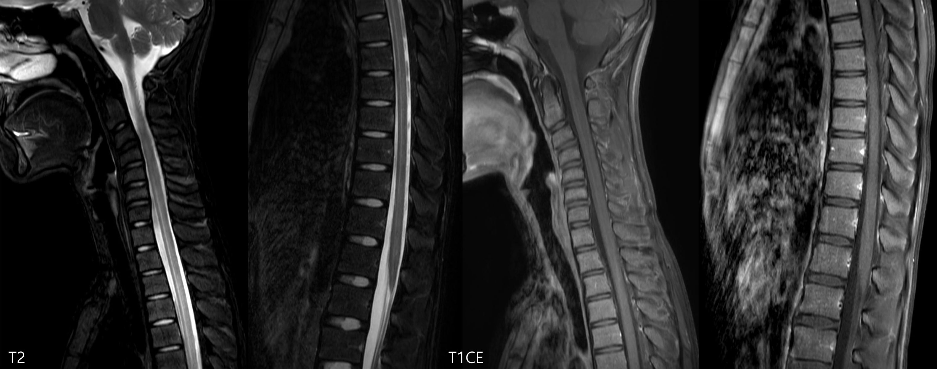
| Clinical information | M/13 Lower limb weakness, urinary retention, ataxia, positive romberg test What is your diagnosis? |
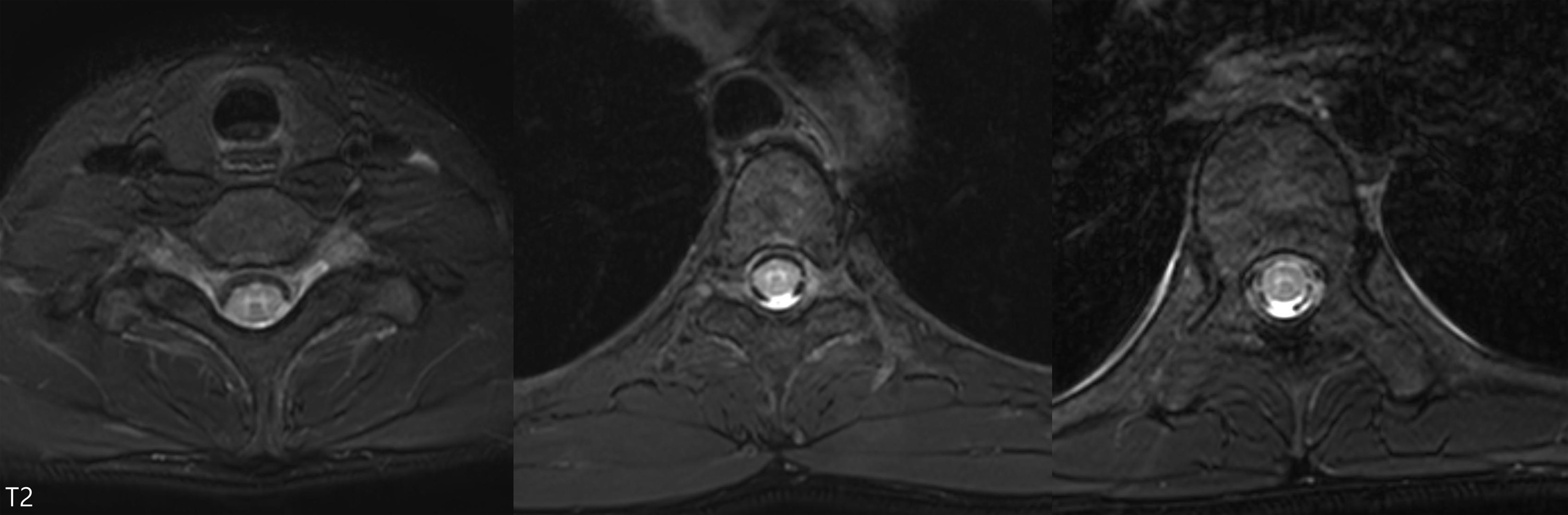
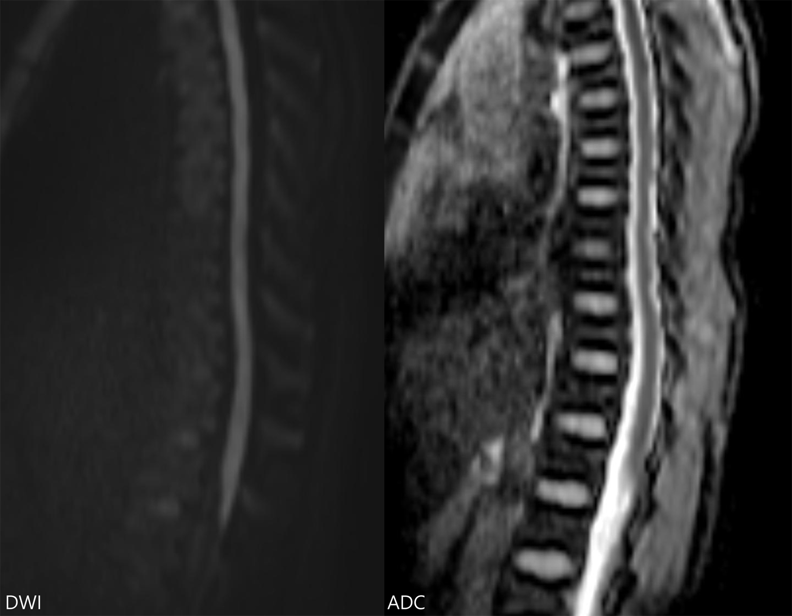
| Discussion | Sagittal T2WI demonstrated a longitudinally extensive hyperintense lesion involving nearly the entire spinal cord. No contrast enhancement was observed. On axial T2WI, the hyperintensity was more prominent in the gray matter. Diffusion-weighted imaging (DWI) showed no evidence of diffusion restriction. The patient tested positive for serum myelin oligodendrocyte glycoprotein (MOG) antibodies and was diagnosed with MOG-associated disease (MOGAD). MOGAD represents a group of inflammatory demyelinating disorders characterized by the presence of IgG antibodies to MOG. Transverse myelitis is the second most common clinical manifestation in MOGAD. Characteristic spinal MRI findings include: Pseudodilatation of the central canal: sagittal T2-hyperintense linear signal surrounded by more hazy T2 hyperintensity in the anterior and posterior gray matter horns H-sign: central T2 hyperintensity on axial imaging, confined to the gray matter, often lacking gadolinium enhancement. Two different patterns depending on the length of involvement: longitudinally extensive transverse myelitis (LETM) or short segment involvement. Contrast enhancement can be observed in some cases Reference) 1. Shahriari M, Sotirchos ES, Newsome SD, Yousem DM. MOGAD: How It Differs From and Resembles Other Neuroinflammatory Disorders. AJR Am J Roentgenol. 2021 Apr;216(4):1031-1039. 2. Zhang L, Feng C, He L, Huang SY, Liu XY, Fan X. MOG-antibody-associated transverse myelitis with the H-sign and unusual MRI enhancement: a case report and literature review. Front Pediatr. 2024 Sep 10;12:1451688. |
| Correct answer |
Correct Answer
Semi-Correct Answer |
Submit answer
Answer submission list
The written answers can only be viewed by the administrator and author.
The written answers can only be viewed by the administrator and author.
The written answers can only be viewed by the administrator and author.