HOME > Weekly Case > Weekly Case
Weekly Case
종결
Case No. 723
2025-07-01~2025-07-31
출제자 : 서배선
Hit : 401
| Period | 2025-07-01~2025-07-31 |
|---|---|
| Diagnosis | Cerebral sparganosis (late stage) |
| Gender |
|
| Age | 51 |
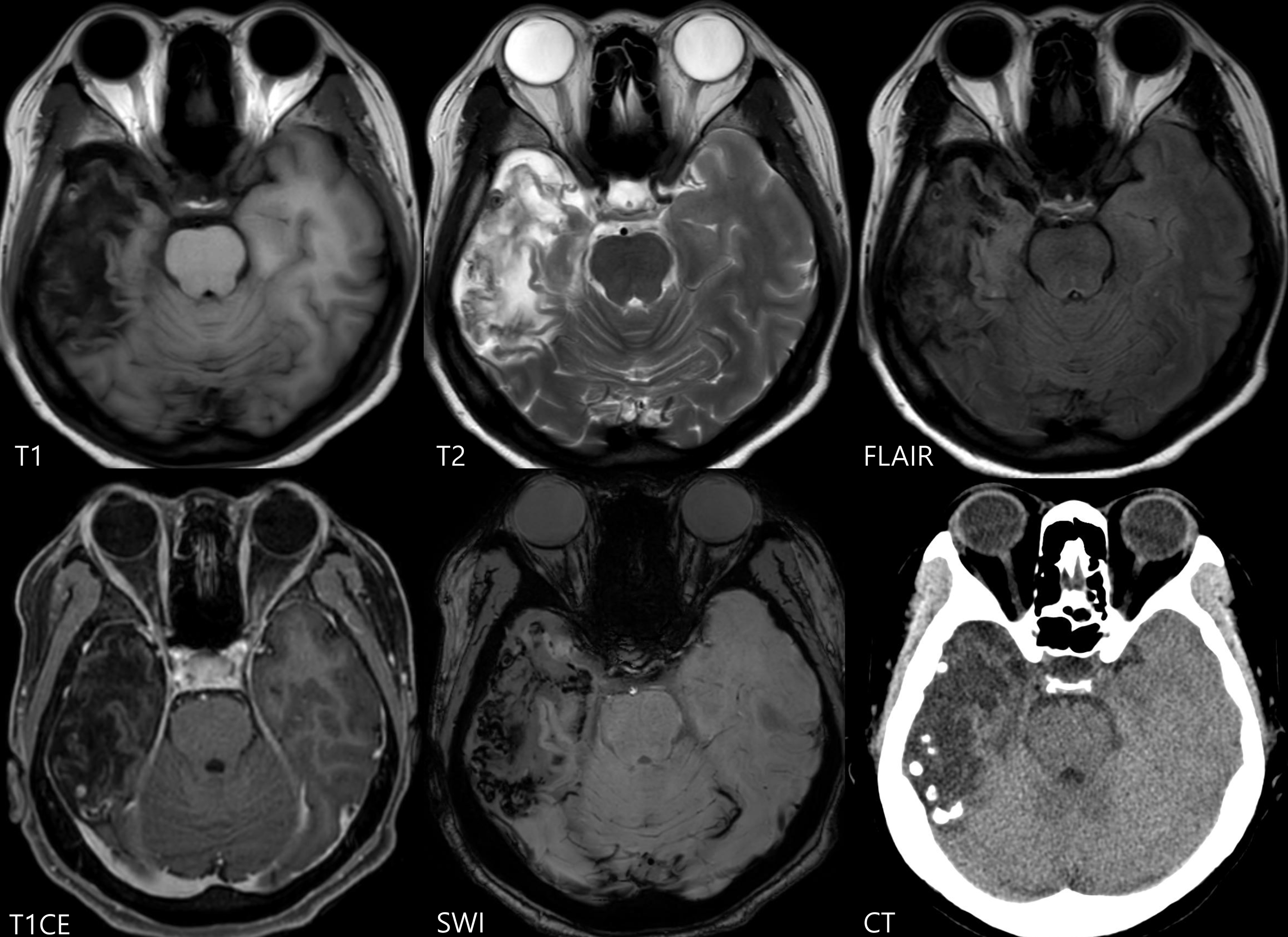
| Clinical information | F/51 Clinical information: Memory impairment since 2016, headache, single seizure episode What is your diagnosis? |
| Discussion | On axial T2-weighted MRI, encephalomalacic changes are observed in the right temporal lobe. Multiple hypointense foci on T2WI and susceptibility-weighted imaging (SWI), consistent with calcifications and petechial hemorrahges, are seen within the encephalomalacic area and are also confirmed on CT. Some of these calcified lesions demonstrate peripheral or tubular contrast enhancement. Although not shown in the provided images, dilatation of the right lateral ventricle is also noted. Serum Sparganum IgG antibodies were positive, and the patient was diagnosed with late-stage cerebral sparganosis. Sparganosis is a rare parasitic infection, most commonly reported in Southeast Asia, China, Japan, and Korea. Human infection typically results from ingesting raw or undercooked meat from infected hosts such as frogs, snakes, or chickens. Cerebral involvement can present with headache, seizures, hemiparesis, or other focal neurological deficits. The most characteristic imaging finding is the "tunnel sign"—a tubular or fusiform, peripherally enhancing lesion on post-contrast MRI—reflecting worm migration with associated granuloma or reactive inflammatory tissue encasing the parasite. Additional enhancement patterns include ring-like or nodular configurations. In the chronic stage, imaging may reveal punctate calcifications around or in the degenerated or dead parasites. Other chronic findings include perilesional white matter edema, encephalomalacia, cortical atrophy, and ipsilateral ventricular dilatation. Reference) Song T, Wang WS, Zhou BR, Mai WW, Li ZZ, Guo HC, Zhou F. CT and MR characteristics of cerebral sparganosis. AJNR Am J Neuroradiol. 2007 Oct;28(9):1700-5. |
| Correct answer |
Correct AnswerSemi-Correct Answer |