HOME > Weekly Case > Weekly Case
Weekly Case
종결
Case No. 714
2025-03-01~2025-03-31
출제자 : 이지영
Hit : 341
| Period | 2025-03-01~2025-03-31 |
|---|---|
| Diagnosis | Intraosseous hemangioma |
| Gender |
|
| Age | 37 |
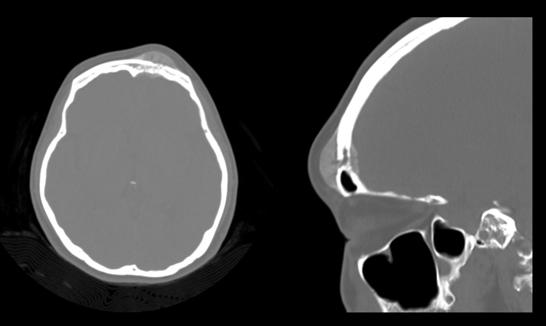
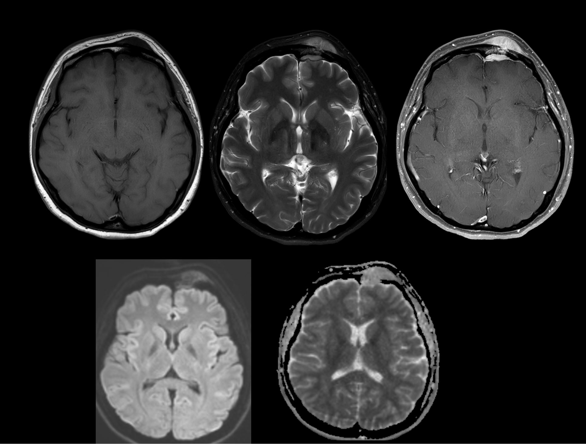
| Clinical information | F/37 c/c: palpable scalp mass on left forehead for 5 months |
| Discussion | -Benign nonmeningothelial mesenchymal bone tumor, most common in the spine -Intracranial hemangiomas Calvarium (m/c), dural venous sinus, dura Peak age 4th-5th decade, M:F ratio 1:2-4 -Clinical presentation: asymptomatic, painless firm mass Slow growing, no malignant transformation -Pathology: Non-encapsulated vascular reddish-brown lesions -Types: Capillary, cavernous (most common), or mixed -Large endothelium-lined spaces separated by fibrous septa -CT Sharply marginated, expansile diploic mass on NECT Inner and outer tables thinned but usually intact Thin sclerotic margin Reticulated hyperdensities from thickened trabeculae → “honeycomb” or “jail bars” appearance -MRI T1WI: Hypo- to isointense with possible scattered hyperintensities due to fat in the lesion T2WI: Markedly hyperintense Post-contrast: Diffuse intense enhancement Dynamic scan: Slow centripetal filling-in |
| Correct answer |
Correct Answer
Semi-Correct Answer |