HOME > Weekly Case > Weekly Case
Weekly Case
종결
Case No. 713
2025-02-01~2025-02-28
출제자 : 최혜정
Hit : 627


| Period | 2025-02-01~2025-02-28 |
|---|---|
| Diagnosis | Merkel cell carcinoma |
| Gender |
|
| Age | 82 |
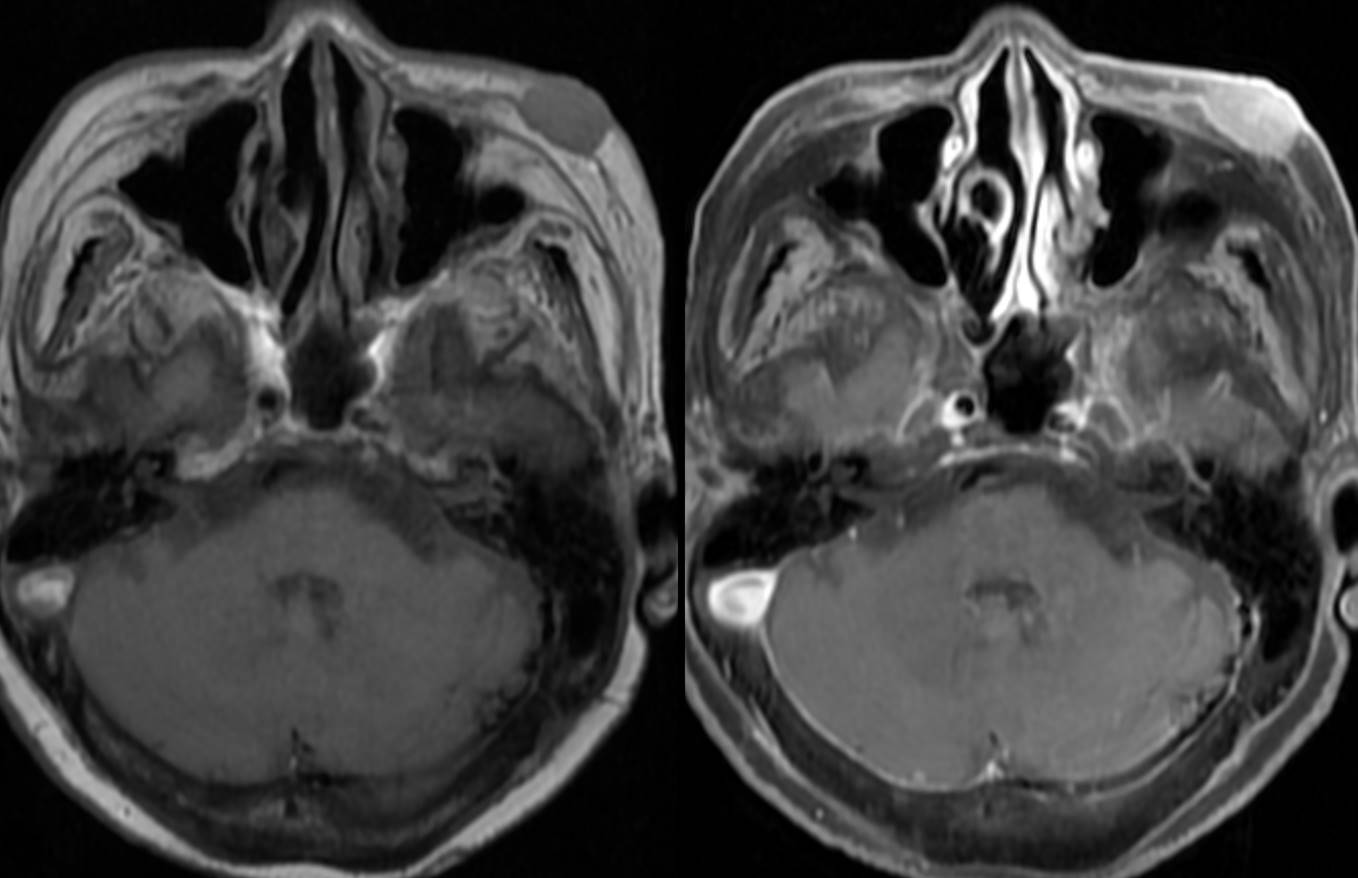
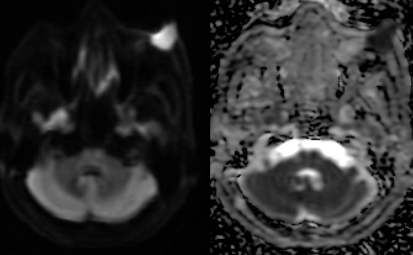
| Clinical information | 82세 여자환자가 rapid growing cheek mass 를 주소로 내원하였다. 진단은? |
| Discussion | Merkel cell carcinoma (MCC) commonly presents as asymptomatic, rapidly growing, dome-shaped nodules, predominantly on the head and neck. MCC typically shows pink to purplish coloration with a shiny or telangiectatic surface, often appearing innocuous in early stages. Key Imaging Findings: 1. Grayscale Sonography: o MCC lesions are generally hypoechoic, with a bilayered appearance in some cases: a superficial, oval, well-defined layer, and a deeper, poorly defined, markedly hypoechoic layer corresponding to subdermal tumor infiltration. o Posterior acoustic enhancement and overlying epidermal thickening were observed in a subset of cases. 2. Color and Power Doppler Sonography: o Most lesions demonstrated rich vascularization with hypertrophic peritumoral and intratumoral vessels. o Vessels often showed a chaotic, irregular vascular architecture, with some exhibiting vertically oriented major vessels. o Spectral analysis in a subset of cases revealed low-velocity, low-resistance arterial flow, a characteristic shared with other soft tissue malignancies. 3. CT : MCC is characterized by areas of high attenuation within the soft tissues, often accompanied by associated lymphadenopathy. Lesions typically demonstrate significant enhancement following contrast administration. 4. MRI : MCC typically appears hypo- to isointense on T1-weighted images and iso- to hyperintense on T2-weighted or fat-saturated T2-weighted images. Following gadolinium administration, the tumor often demonstrates diffuse or heterogeneous enhancement. Larger lesions may exhibit inhomogeneous signal intensity on both T1- and T2-weighted sequences, with areas of focal central hyperintensity on T2-weighted images, indicative of necrosis or hemorrhage. 5. PET : FDG-avid, hypermetabolic lesion, reflecting its aggressive nature. Treatment and Prognosis: The primary treatment for localized MCC involves surgical excision of the tumor, often combined with adjuvant radiotherapy. For advanced or refractory cases, immune checkpoint inhibitors have shown promise as an effective therapeutic option. Reference 1. Orlando Catalano et al. Color Doppler Sonography of Merkel Cell Carcinoma J Ultrasound Med. 2018 Jan;37(1):285-292. doi: 10.1002/jum.14329. Epub 2017 Jul 20. 2. Akaike, G., Akaike, T., Fadl, S. A., Lachance, K., Nghiem, P., & Behnia, F. (2019). Imaging of Merkel Cell Carcinoma: What Imaging Experts Should Know. RadioGraphics, 39(7), 2069–2084. doi:10.1148/rg.2019190102 |
| Correct answer |
Correct Answer
Semi-Correct Answer |