HOME > Weekly Case > Weekly Case
Weekly Case
종결
Case No. 710
2025-01-01~2025-01-31
출제자 : 박상익
Hit : 661
| Period | 2025-01-01~2025-01-31 |
|---|---|
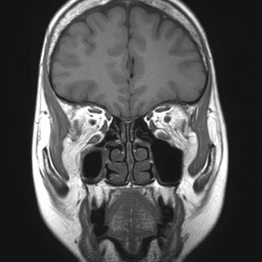
| Diagnosis | Orbital fat prolapse through inferior orbital fissure |
| Gender |
|
| Age | 63 |
| Clinical information | 1개월 전부터 시작된 복시를 주소로 내원한 63세 여자 환자이다. 진단은? |
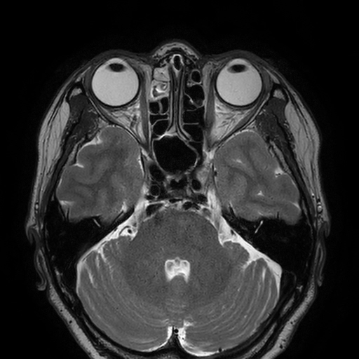
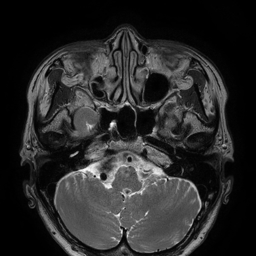
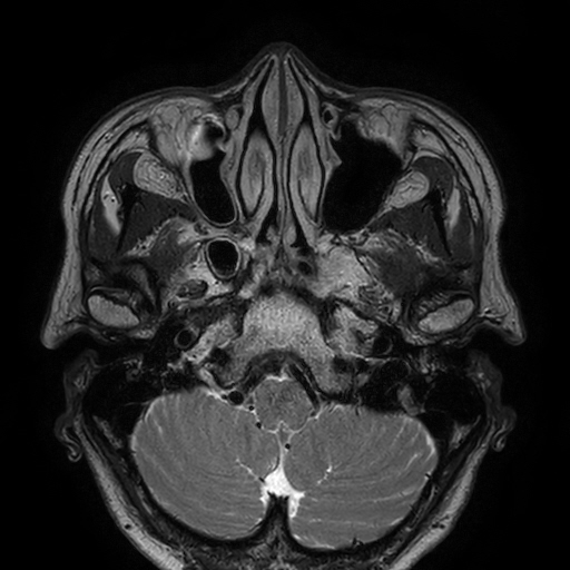
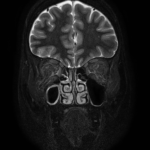
| Discussion | On axial T2-weighted images, heterogeneously T2-hyperintense structures are seen abutting the maxillary sinus walls in the bilateral infratemporal fossae, appearing to connect to the orbits via the inferior orbital fissures. Coronal STIR and T1-weighted images show inferolateral herniation of bilateral orbital fat into the infratemporal fossae, along with infiltrations within the intraorbital fat and prolapsed fat. Orbital fat volumes appear decreased, and bilateral enophthalmos is suspected on MRI. Orbital fat prolapse through the inferior orbital fissure is not an uncommon finding, with a reported prevalence of 9% (20/228) in an orbital MRI study. It was unilateral in 55% of patients and bilateral in 45%, with no significant association found with age, sex, obesity, Graves disease, hypercortisolism, prior orbital trauma, proptosis, or enophthalmos. In this patient, the orbital fat prolapse was considered pathological by the reporting radiologist due to the degree of herniation and the presence of fat infiltrations. However, the patient tested positive for acetylcholine receptor antibodies and was clinically diagnosed with ocular myasthenia gravis. Her symptoms showed some improvement after starting pyridostigmine. The extent to which orbital fat prolapse contributed to her symptoms remains uncertain. Reference 1. Bunch PM, Buch K, Kelly HR. Prolapse of Orbital Fat through the Inferior Orbital Fissure: Description, Prevalence, and Assessment of Possible Pathologic Associations. AJNR Am J Neuroradiol. 2019;40(8):1388-91. |
| Correct answer |
Correct Answer
Semi-Correct Answer |