HOME > Weekly Case > Weekly Case
Weekly Case
종결
Case No. 708
2025-01-01~2025-01-31
출제자 : 박상익
Hit : 370
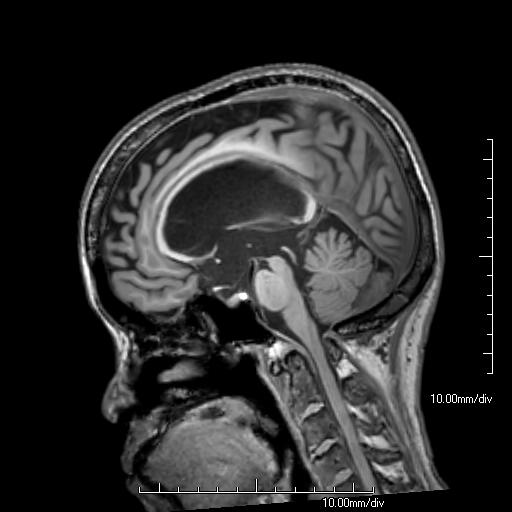
| Period | 2025-01-01~2025-01-31 |
|---|---|
| Diagnosis | Achondroplasia |
| Gender |
|
| Age | 61 |
| Clinical information | 수 년 전부터 진행하는 기억장애를 주소로 내원한 61세 여자 환자이다. 이 환자의 기저 질환은? |
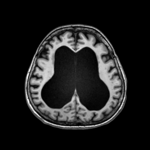
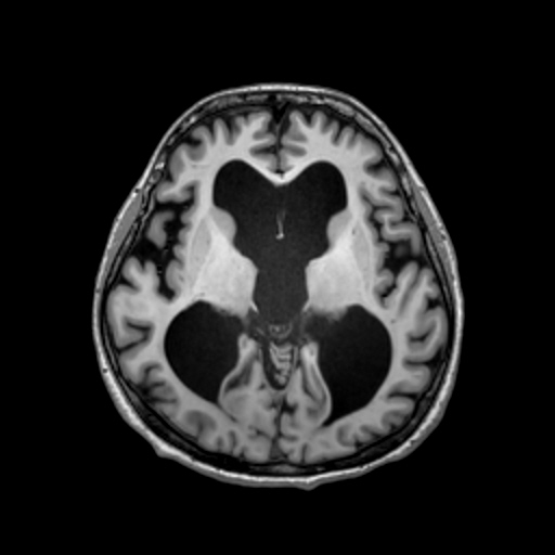
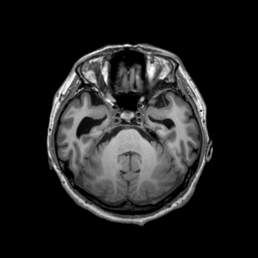
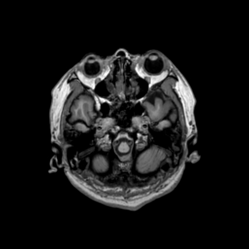
| Discussion | On axial T1-weighted images, there is enlargement of the lateral and third ventricles with a relatively normal-sized fourth ventricle. The sagittal T1-weighted image does not show aqueductal stenosis. The skull base is hypoplastic, with a hypoplastic clivus and narrowing of the foramen magnum. Additionally, there is moderate, diffuse central canal stenosis in the upper cervical spine, as observed on the sagittal T1-weighted image. These findings are consistent with the cranial and spinal manifestations of achondroplasia, an autosomal dominant condition caused by pathogenic variants in the fibroblast growth factor receptor 3 (FGFR3) gene. The cranial features of achondroplasia include macrocephaly with a relatively small skull base and a narrow foramen magnum. The characteristic pattern of hydrocephalus in patients with achondroplasia is “triventricular,” with crowding of the posterior fossa but without stenosis of the cerebral aqueduct, as demonstrated in this patient. This type of hydrocephalus is generally considered to be communicating hydrocephalus, with increased venous sinus pressure likely secondary to jugular foramen stenosis. References 1. Campbell J, Legare JM, Piatt J, Gough E, Pauli RM, Hashmi SS, et al. Achondroplasia Natural History Study (CLARITY): 60-year experience with hydrocephalus in achondroplasia from four skeletal dysplasia centers. J Neurosurg Pediatr. 2023;32(6):649-56. 2. Kim J, Patel VJ, El Ahmadieh TY, Olson DM, Swift DM. Hydrocephalus in achondroplasia: efficacy of endoscopic third ventriculostomy. J Neurosurg Pediatr. 2022;29(3):268-75. |
| Correct answer |
Correct Answer
Semi-Correct Answer |