HOME > Weekly Case > Weekly Case
Weekly Case
종결
Case No. 701
2024-10-01~2024-11-30
출제자 : 박미나
Hit : 420
| Period | 2024-10-01~2024-11-30 |
|---|---|
| Diagnosis | intramedullary melanoma |
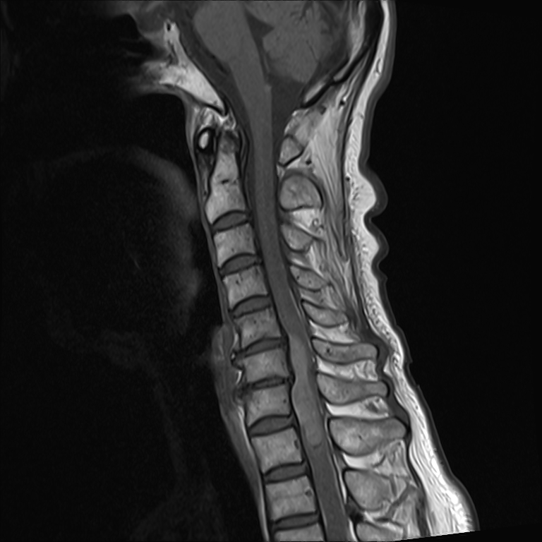
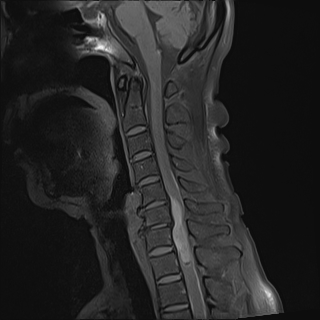
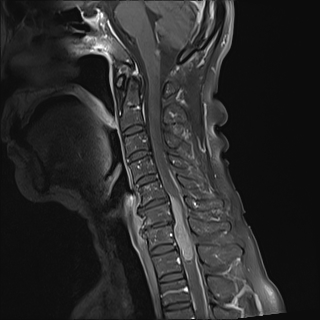
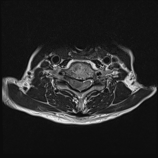
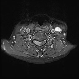
| Clinical information | 우연히 발견된 척수내 종양이다. 진단은? |
| Discussion | 진단: intramedullary melanoma Primary melanoma of the central nervous system (CNS) is a rare neoplasm that accounts for approximately 1% of all cases of melanoma. Primary intramedullary spinal cord melanoma is an extremely unique entity most of its cases seem to arise in the thoracic spinal cord. Location of spinal tumors can be intra- or extramedullary, leptomeningeal, or extradural. Intramedullary location. MRI of primary CNS melanoma shows hyperintense signals on T1-weighted imaging and hypointense signals on T2- and T2*-weighted imaging. This depends on the degree of pigment content. Enhancement patterns may also be inhomogeneous, peripheral, or nodular. Lesions secondary to leptomeningeal dissemination also display hyperintense signals on T1-weighted imaging, with gadolinium enhancement and hypointense signals on T2-weighted imaging. |
| Correct answer |
Correct Answer
Semi-Correct Answer |