HOME > Weekly Case > Weekly Case
Weekly Case
종결
Case No. 698
2024-09-01~2024-09-30
출제자 : 조세진
Hit : 824
| Period | 2024-09-01~2024-09-30 |
|---|---|
| Diagnosis | Juvenile/Adult-onset Alexander disease |
| Gender |
|
| Age | 21 |
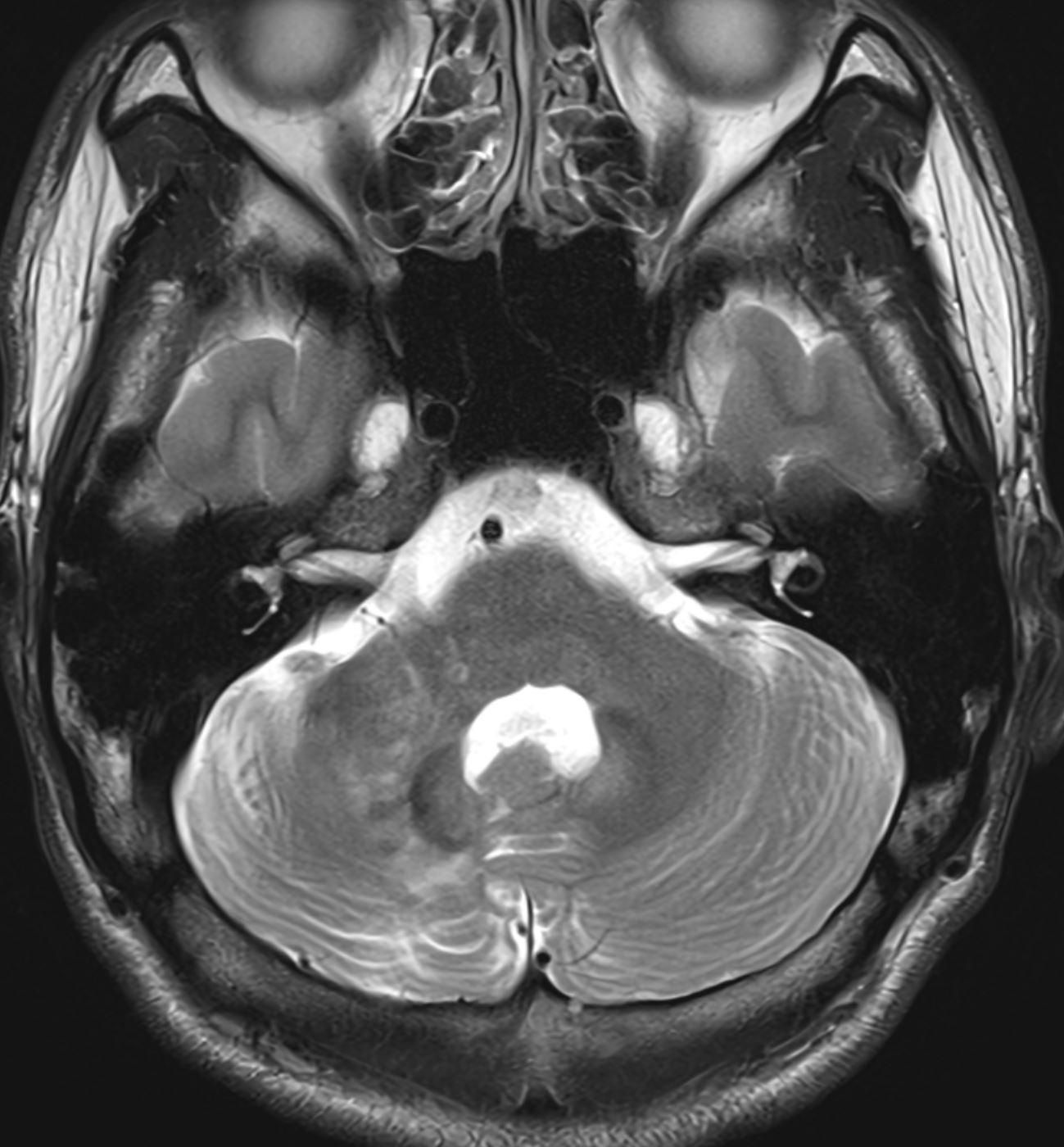
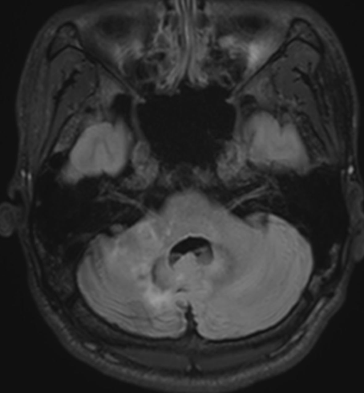
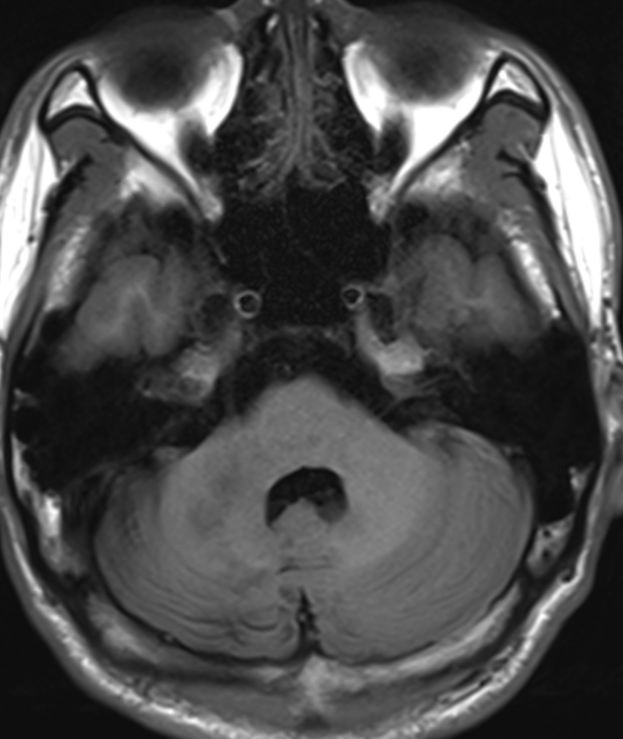
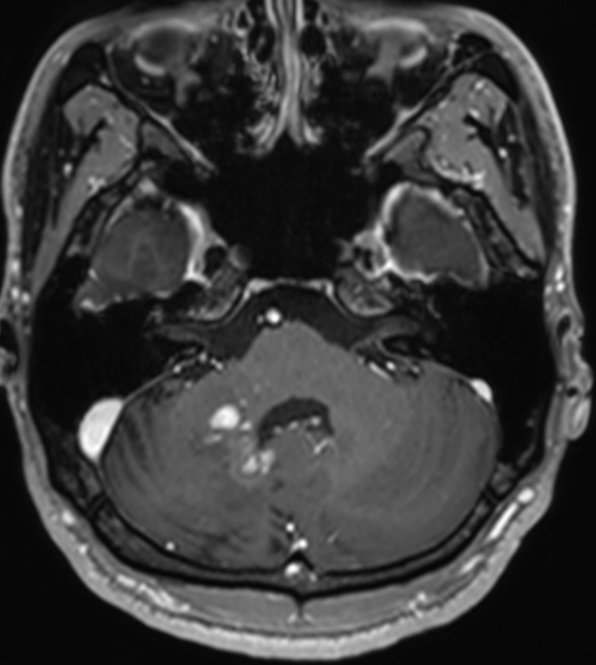
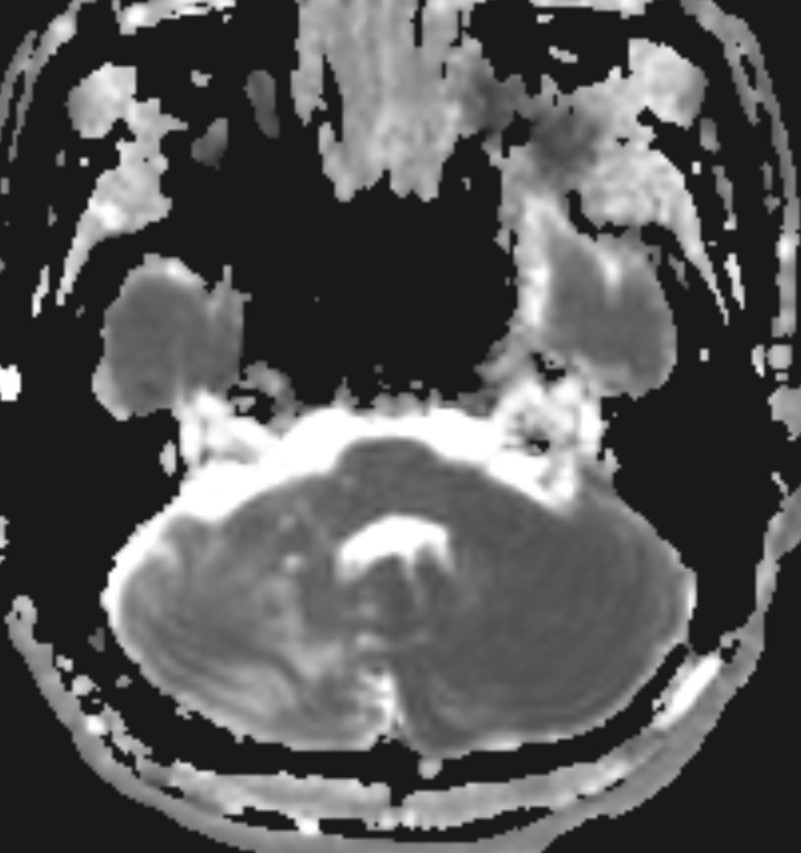
| Clinical information | M/21Y, 반복적인 우측 ataxia 와 dysmetria로 내원한 환자가 보인 소뇌 병변, 진단은? Images> T2WI, FLAIR, T1WI, contrast T1WI, ADC 분자유전학검사> 본 환자의 GFAP 유전자에서 기보고된 likely pathogenic variant인 c.209G>A, p.Arg70Gln, heterozygote가 검출되었습니다. |
| Discussion | 진단: Juvenile/Adult-onset Alexander disease Review Alexander disease (AD) was known as a rare, genetically determined leukoencephalopathy affecting infants and children, characterized by macrocephaly, seizures, spasticity, and retarded psychomotor development, and leading to death in 2 months to 7 years.1 The diagnostic MR imaging features established by van der Knaap et al consisted of extensive white matter (WM) increased signal intensity on T2-weighted images, mainly in the frontal regions; a rim of periventricular T2 hypointensity; involvement of the basal ganglia, thalami, and brain stem; and postcontrast enhancement in the periventricular regions and scattered areas of the brain stem. These features allowed the diagnosis without biopsy, which was performed in the search for Rosenthal fibers, the histologic hallmark of the disease. Milder forms with spastic paraparesis, ataxia, or lower brain stem signs and juvenile (2–12 years of age) or adult onset (≥13 years) had been diagnosed as AD at postmortem examination because of the presence of Rosenthal fibers.4 After 2001, the discovery that mutations in the glial fibrillary acidic protein gene (GFAP) were responsible for the disease5,6 allowed recognition during life of juvenile or adult forms1,7–20 that have an MR imaging pattern remarkably different from that observed in children, with predominant focal involvement of the lower brain stem. Reference: Amstutz DR, Coons SW, Kerrigan JF et-al. Hypothalamic hamartomas: Correlation of MR imaging and spectroscopic findings with tumor glial content. AJNR Am J Neuroradiol. 2006;27 (4): 794-8. AJNR Am J Neuroradiol (full text) - Pubmed citation |
| Correct answer |
Correct Answer
Semi-Correct Answer |