HOME > Weekly Case > Weekly Case
Weekly Case
종결
Case No. 695
2024-08-01~2024-08-31
출제자 : 황인평
Hit : 508
| Period | 2024-08-01~2024-08-31 |
|---|---|
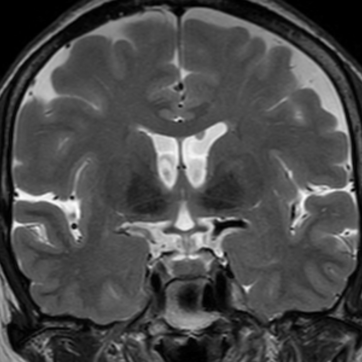
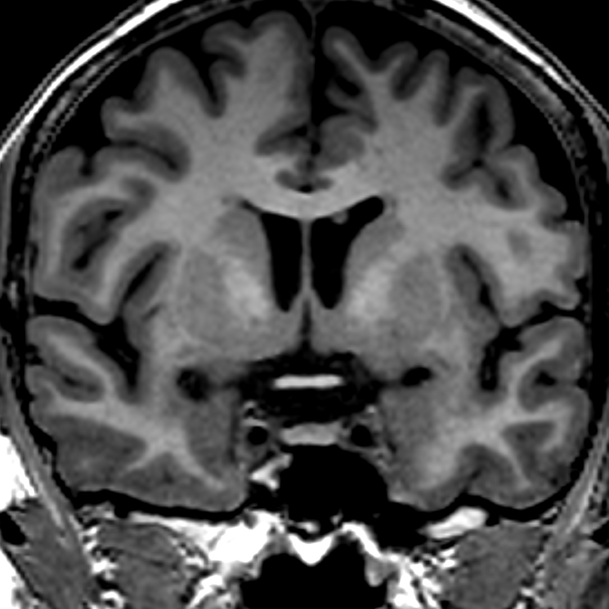
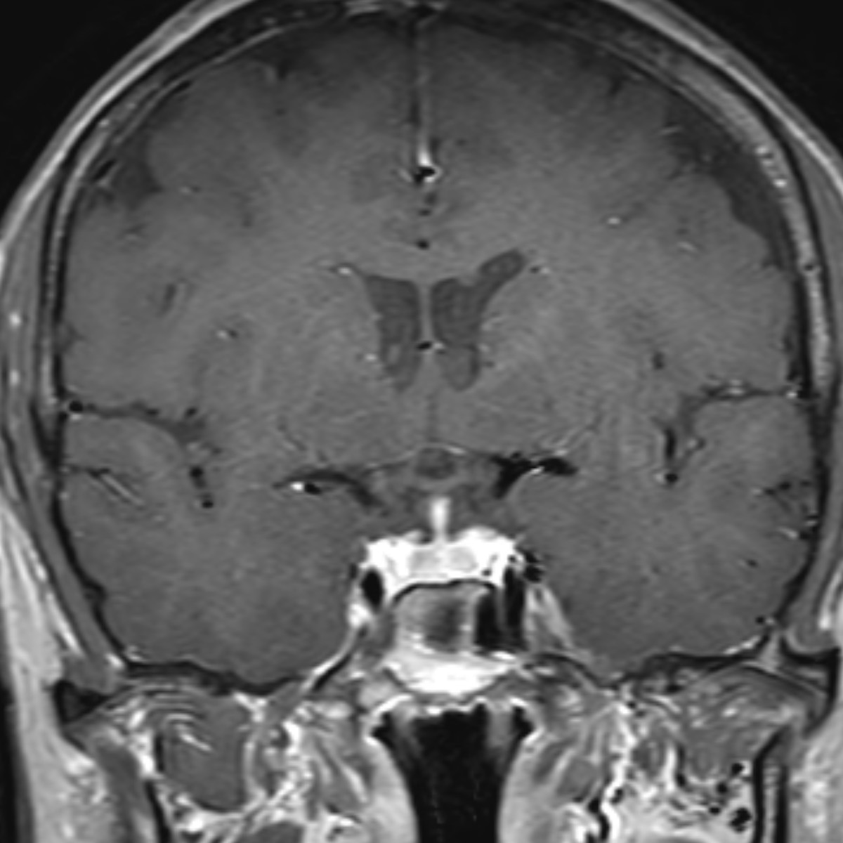
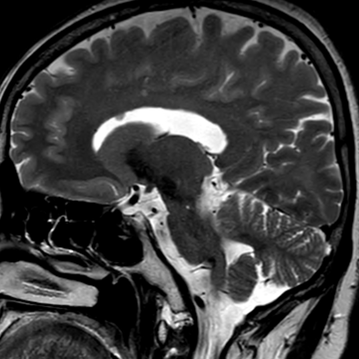
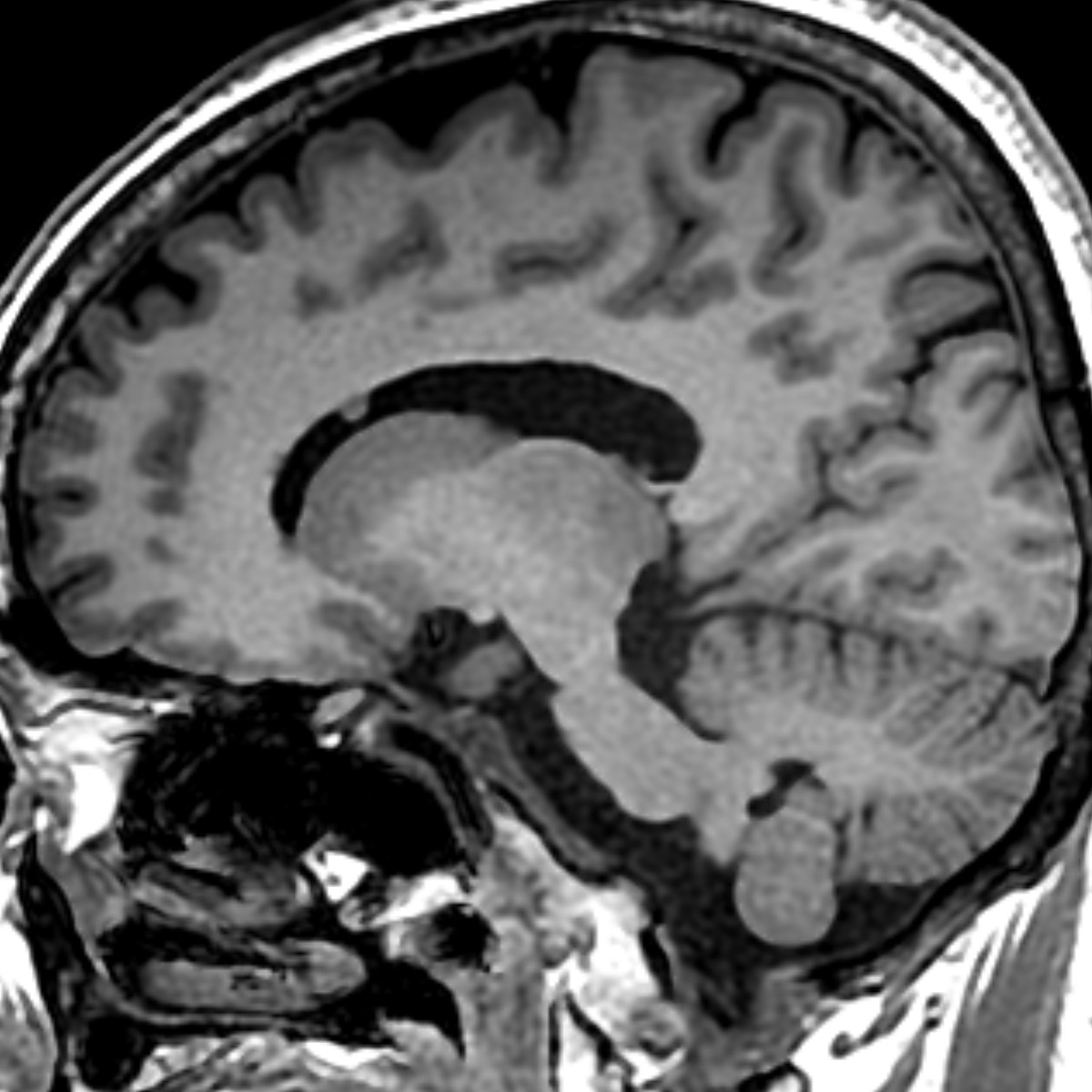
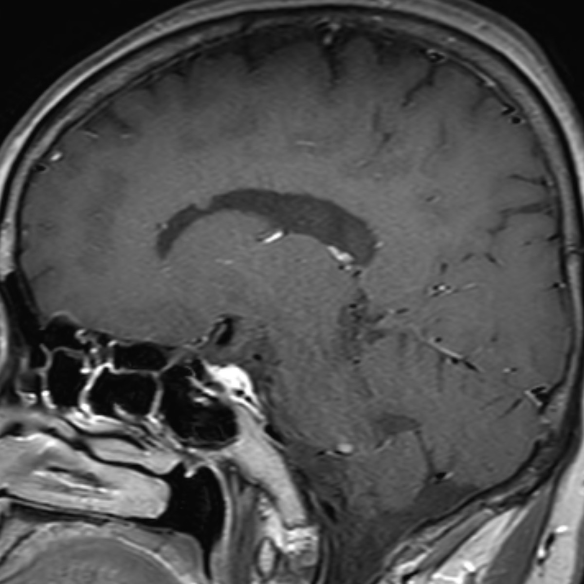
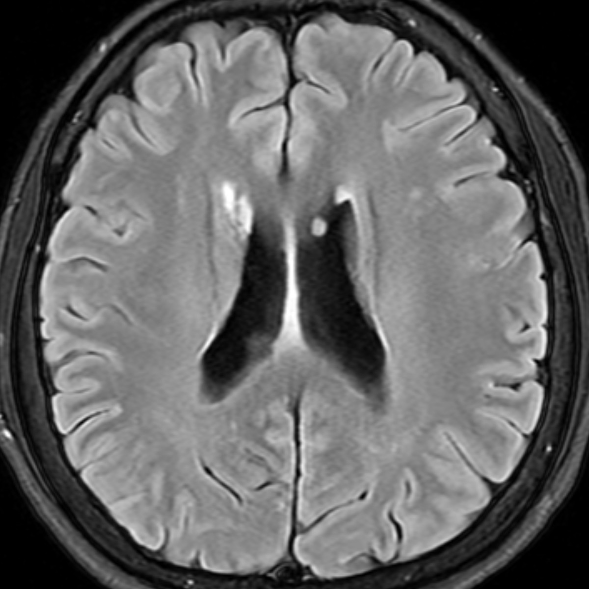
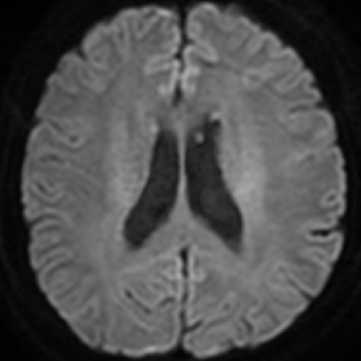
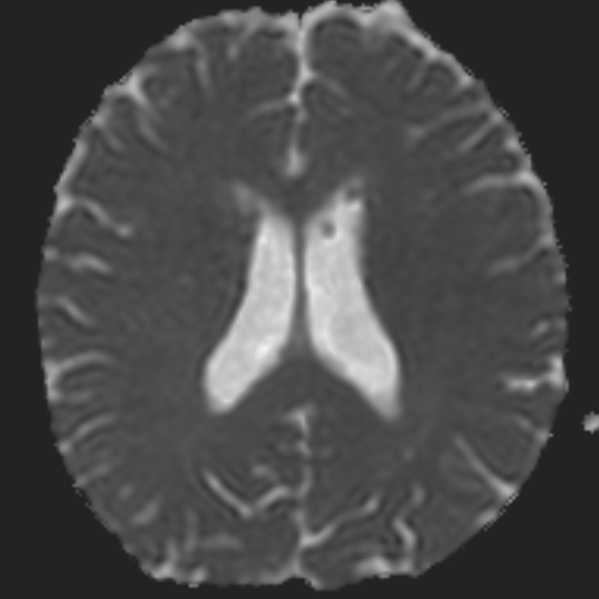
| Diagnosis | Ring-shaped lateral ventricular nodules |
| Gender |
|
| Age | 58 |
| Clinical information | 56세 여성 환자입니다. 뇌실안에 보이는 병변은 무엇일까요? |
| Discussion | Ring-shaped lateral ventricular nodules Incidentally encountered lesions, <1 cm, 0.023% Several hypotheses: neuroglial/glioependymal cyst, inflammatory or reactive formation of the ependyma, astrocytic gliosis reaction near subependymal veins, redetachment of the fused portion of coarctation of the frontal horn, or a variant of subependymoma Imaging: T1w, T2w: isointesnse to gray matter FLAIR: iso to hyeprintense No contrast enhancement No rapid growth Shared similar anatomical location: occurring in the roof of body or frontal horn of the lateral ventricles DDx.: subependymal heterotopias, subependymal nodules (tuberous sclerosis), subependymoma, subependymal giant cell astrocytoma. Reference Shimono T et al. Neuroradiology. 2009;51(3):145-50. Pontillo G et al. World Neurosurg. 2020;136:32-36. |
| Correct answer |
Correct Answer
Semi-Correct Answer |