HOME > Weekly Case > Weekly Case
Weekly Case
종결
Case No. 687
2024-06-01~2024-06-30
출제자 : 이경식
Hit : 337
| Period | 2024-06-01~2024-06-30 |
|---|---|
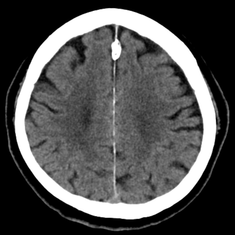
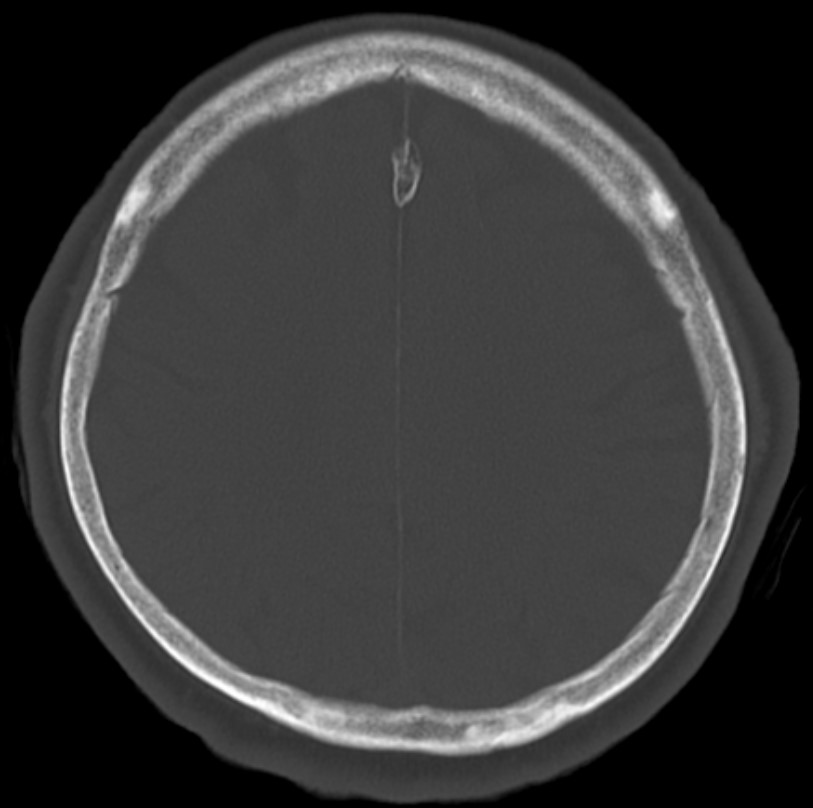
| Diagnosis | falx ossification |
| Gender |
|
| Age | 82 |
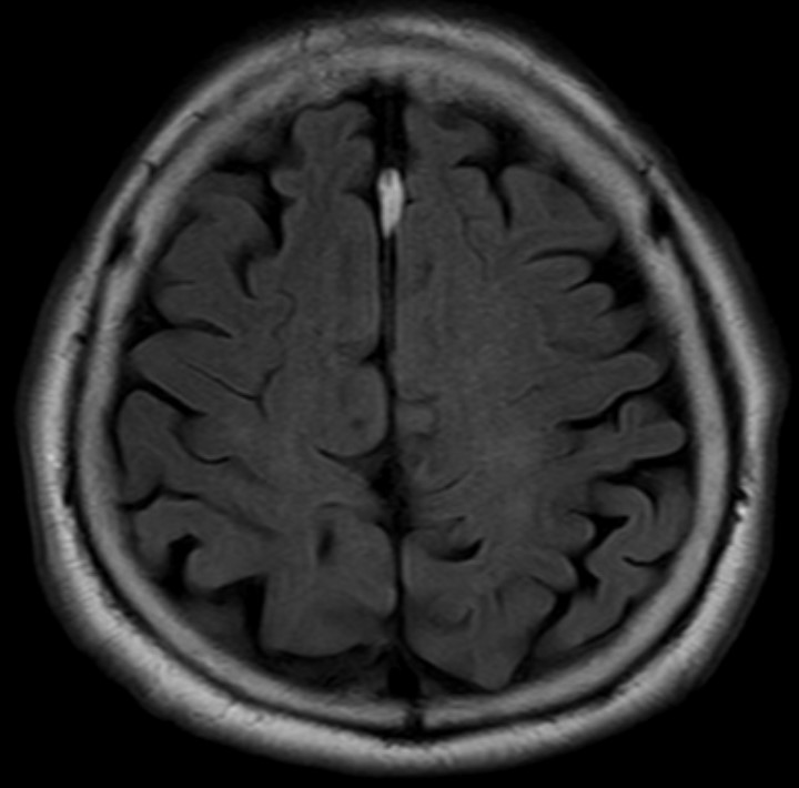
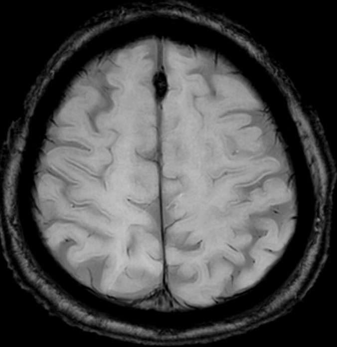
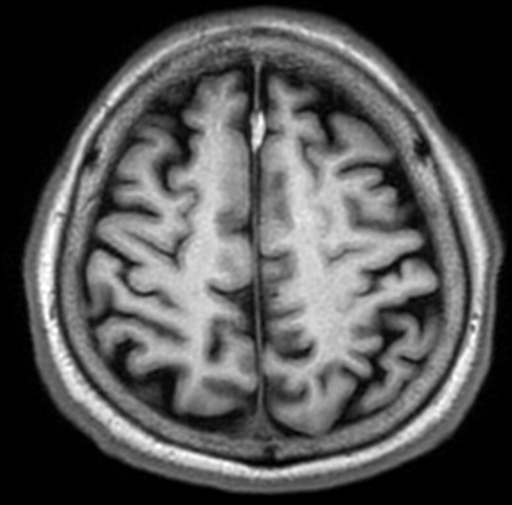
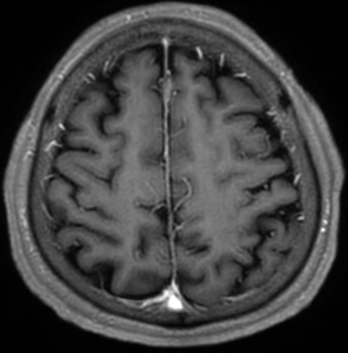
| Clinical information | 외상 및 일시적 의식 소실이 있는 82세 여자의 CT와 MRI 영상입니다. 가능한 진단은? |
| Discussion | Falx cerebri가 focal mineralization이 된 것으로, 단순한 falx calcification과는 다르며, incidence는 매우 낮은 것으로 보고되고 있다. falx cerebri은 mesenchyma에서 유래하기 때문에, 골화를 유발하는 요인들 (마찰, 출혈 또는 외상등)로 인해 골형성 변화 및 막뼈를 형성하게 된다고 알려져 있다. Imaging feature: 1) CT: isolated ossification of the falx 2) MRI: typical appearance consisting of a central marrow-containing portion with signal intensity similar to fat (bright on T1), surrounded by low signal intensity representing cortical bone DDx: fatty falx cerebri, falx cerebri calcification, focal SDH |
| Correct answer |
Correct Answer
Semi-Correct Answer |