HOME > Weekly Case > Weekly Case
Weekly Case
종결
Case No. 686
2024-04-01~2024-04-30
출제자 : 임영희
Hit : 899
| Period | 2024-04-01~2024-04-30 |
|---|---|
| Diagnosis | venous infarction |
| Gender |
|
| Age | 65 |
| Clinical information | 65M cognitive impairment 진단은? |
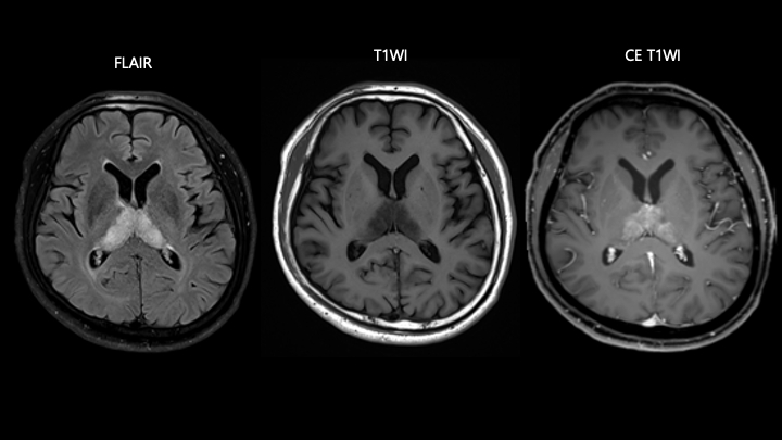
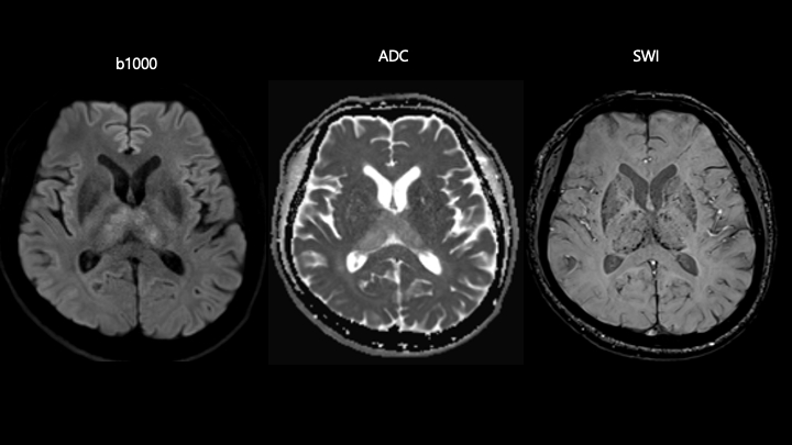
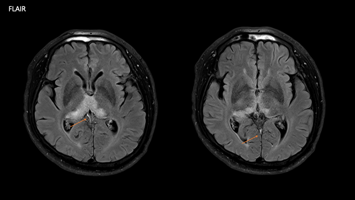
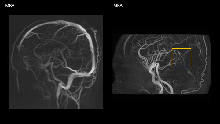
| Discussion | 1. Bilateral T2/FLAIR high SI lesions in the both thalami - petechial hemorrhages in the bilateral thalami and corpus callosum/cingulate gyri 2. Loss of flow void in the straight sinus and internal cerebral veins on FLAIR; invisible straight sinus on MRV and prominent vascular structures around the straight sinus on MRA. Venous infarctions resulted from chronic venous sinus thrombosis, R/O combined with dural AVF Bithalamic lesions Cause Differential Vascular Deep cerebral vein thrombosis and dural venous sinus thrombosis Internal cerebral veins Vein of Galen Straight sinus Arterial infarction Artery of Percheron occlusion top of the basilar artery syndrome PRES (Posterior reversible encephalopathy syndrome) Infection CJD Viral encephalitis (West Nile encephalitis, Japanese encephalitis, Murray Valley encephalitis, Eastern equine encephalitis, and rabies) Malaria Metabolic and Toxic Disorders Wernicke Encephalopathy Osmotic Myelinolysis Fabry Disease Fahr Disease Wilson Disease Leigh Disease Deep venous thrombosis Typically results in bilateral symmetric involvement of thalami and occasionally basal ganglia. Causes include pregnancy, oral contraceptives, infection, trauma, and dehydration, undetermined in 20~25%. Infarct or hemorrhage in atypical areas crossing arterial territories or infarct with cortical sparing Reference AJR, Volume 192, Issue 2, February 2009, https://doi.org/10.2214/AJR.08.1585 |
| Correct answer |
Correct Answer
Semi-Correct Answer |