HOME > Weekly Case > Weekly Case
Weekly Case
종결
Case No. 678
2024-01-01~2024-01-31
출제자 : 서종현
Hit : 578
| Period | 2024-01-01~2024-01-31 |
|---|---|
| Diagnosis | Neuronal intranuclear inclusion disease (NIID) |
| Gender |
|
| Age | 76 |
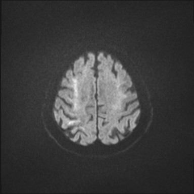
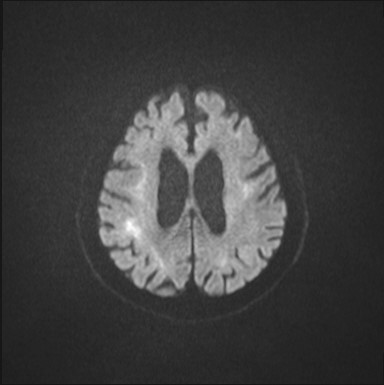
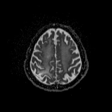
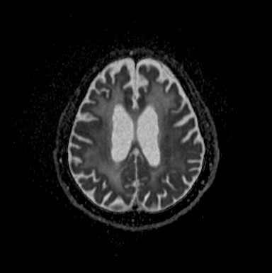
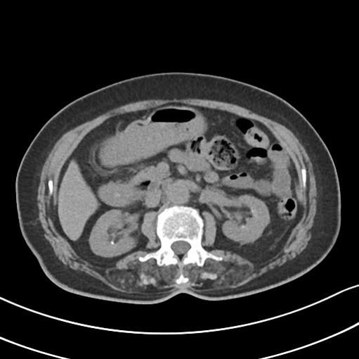
| Clinical information | 기억력 장애로 내원한 76세 여자환자이다. 진단은? |
| Discussion | Neuronal intranuclear inclusion disease (NIID) Skin, ( abdomen ), punch biopsy: - A few P62-positive intranuclear and cytoplasmic inclusions in peripheral nerve and skin adnexa, suggestive of neuronal intranuclear inclusion disease. 1. Introduction Neuronal intranuclear hyaline inclusion disease (NIID) is a slowly progressive neurodegenerative disease characterized by eosinophilic hyaline intranuclear inclusions in the central and peripheral nervous system, and also in the visceral organs. 2. Radiologic findings Conventional brain MRI findings of patients with neuronal intranuclear hyaline inclusion disease strongly resemble those seen in fragile X-associated tremor/ataxia syndrome (FXTAS), including symmetric white matter involvement in combination with hyperintense changes of the middle cerebellar peduncles. Although not invariably present, only NIID typically presents on DWI with highly characteristic band-like and symmetric hyperintense changes of the corticomedullary junctions. This imaging feature is considered a strong indicator of NIID, while symmetric high signal changes on T2/FLAIR in the cerebellar hemispheres and along the cerebellar vermis are occasional ancillary findings. Cortical contrast enhancement with corresponding edema and high signal on DWI has been found in a subset of patients with a younger age of onset, shorter duration of disease, and a higher incidence of a headache than those without enhancement. The enhanced lesions were selectively spread along the surface of posterior cortex and were clinically associated with encephalopathy-like episodes. |
| Correct answer |
Correct Answer
Semi-Correct Answer |