HOME > Weekly Case > Weekly Case
Weekly Case
종결
Case No. 671
2023-10-01~2023-10-31
출제자 : 조세진
Hit : 607
| Period | 2023-10-01~2023-10-31 |
|---|---|
| Diagnosis | Hypothalamic hamartoma |
| Gender |
|
| Age | 40 |
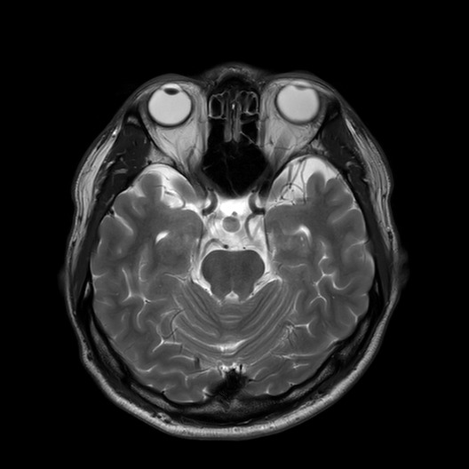
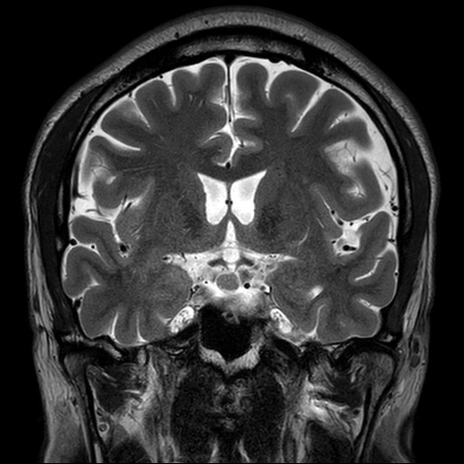
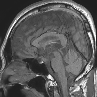
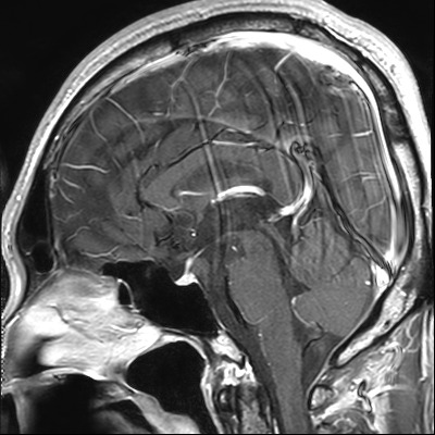
| Clinical information | Question> 건강검진상에서 보인 두개강 내 이상 소견으로 내원, 진단은? M,/40Y Symptom: none Images> Axial & coronal T2WI, sagittal non-contrast & contrast T1WIs |
| Discussion | Review Hypothalamic hamartomas, also known as tuber cinereum hamartomas, are benign non-neoplastic heterotopias in the brain that typically occur in the region of the hypothalamus, arising from the tuber cinereum, a part of the hypothalamus located between the mammillary bodies and the optic chiasm Imaging findings: CT: soft tissue nodule of similar attenuation to the adjacent brain, without calcification or contrast enhancement. Occasionally, it may show suprasellar calcifications, an eroded dorsum sellae, or enlarged pituitary fossa. However, the diagnosis can be challenging, depending on slice position and thickness, although with more recent volumetric scans with coronal and sagittal reformats, the abnormality is easier to identify. MRI: T1: hypointense to the cerebral cortex T1 C+ (Gd): no contrast enhancement T2 hyperintense to the cerebral cortex Reference: Amstutz DR, Coons SW, Kerrigan JF et-al. Hypothalamic hamartomas: Correlation of MR imaging and spectroscopic findings with tumor glial content. AJNR Am J Neuroradiol. 2006;27 (4): 794-8. AJNR Am J Neuroradiol (full text) - Pubmed citation |
| Correct answer |
Correct Answer
Semi-Correct Answer |