HOME > Weekly Case > Weekly Case
Weekly Case
종결
Case No. 669
2023-10-01~2023-10-31
출제자 : 조세진
Hit : 360
| Period | 2023-10-01~2023-10-31 |
|---|---|
| Diagnosis | Giant parietal foramina or Potocki-Shaffer syndrome |
| Gender |
|
| Age | 17 |
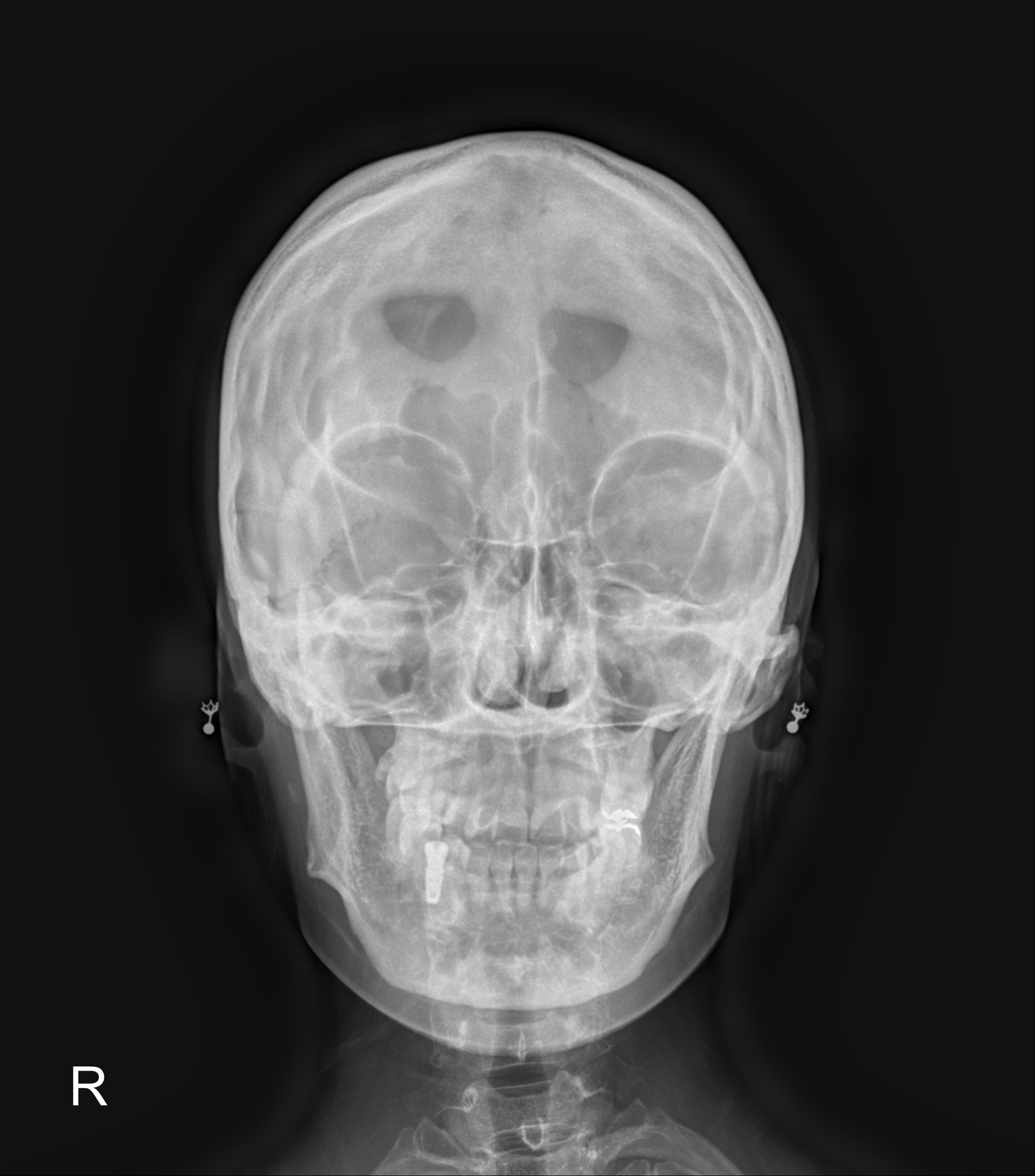
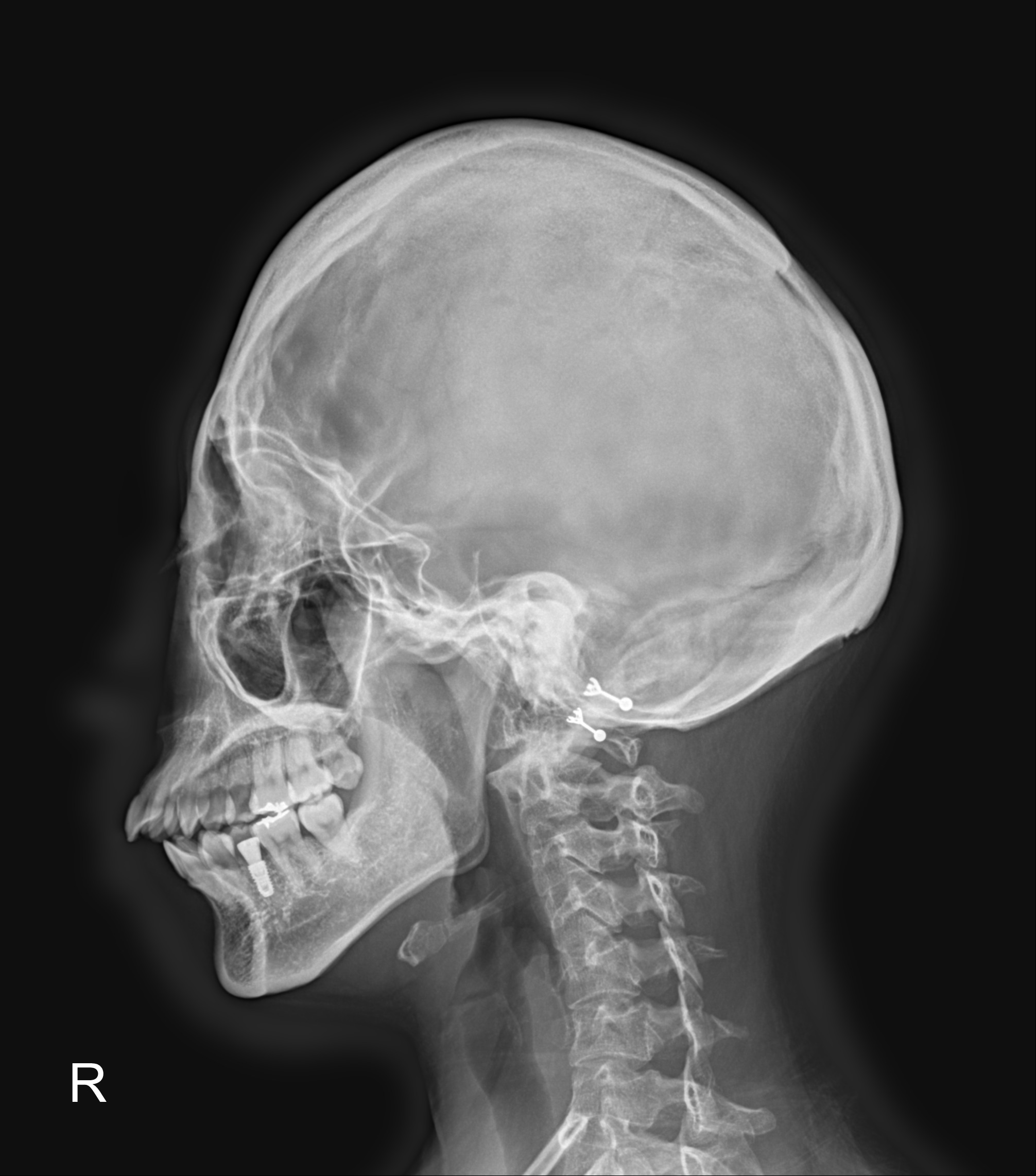
| Clinical information | Question> 무증상 환자, 교통사고로 시행한 외부 검사에서 이상 소견 발견하여 내원한 17세 여자 환자의 영상이다. 1) 가능한 판독과 2)연관이 있다고 알려진 질환은? Images> AP, Lateral skull radiograph |
| Discussion | Increased incidence of venous and cortical anomalies 4. Bilateral parietal foramina are associated with Potocki-Shaffer syndrome. They can occur as an isolated autosomal dominant trait or as part of a syndrome. Ossification along a midline bar may separate confluent parietal defects into paired parasagittal defects during the first few months of life, which can persist into adult life. Giant parietal foramina are thought to be pathologically distinct from the small parietal foramina found in many individuals which transmit emissary veins Underlying genetic anomalies have been identified in chromosome 11p deletions with mutation of the ALX4 (60%) and MSX2 (40%) genes in those with enlarged parietal foramina. Imaging findings: Fetal diagnosis: Second trimester ultrasound (or MRI) may identify the calvarial defects. CT: Seen as paired, rounded defects in the parietal bone located near the intersection of the sagittal and lambdoid sutures. The defects may be large and confluent across the midline. MRI Superior to CT for detecting any associated cortical, venous or meningeal anomalies. Reference: Chung HY, Uster-friedberg T, Pentaz S et-al. Enlarged parietal foramina: findings on prenatal ultrasound and magnetic resonance imaging. Ultrasound Obstet Gynecol. 2010;36 (4): 521-2. doi:10.1002/uog.7731 - Pubmed citation Reddy AT, Hedlund GL, Percy AK. Enlarged parietal foramina: association with cerebral venous and cortical anomalies. Neurology. 2000;54 (5): 1175-8. Neurology (full text) - Pubmed citation |
| Correct answer |
Correct Answer
Semi-Correct Answer |