HOME > Weekly Case > Weekly Case
Weekly Case
종결
Case No. 666
2023-09-01~2023-09-30
출제자 : 박미나
Hit : 404
| Period | 2023-09-01~2023-09-30 |
|---|---|
| Diagnosis | Cerebral fat embolism |
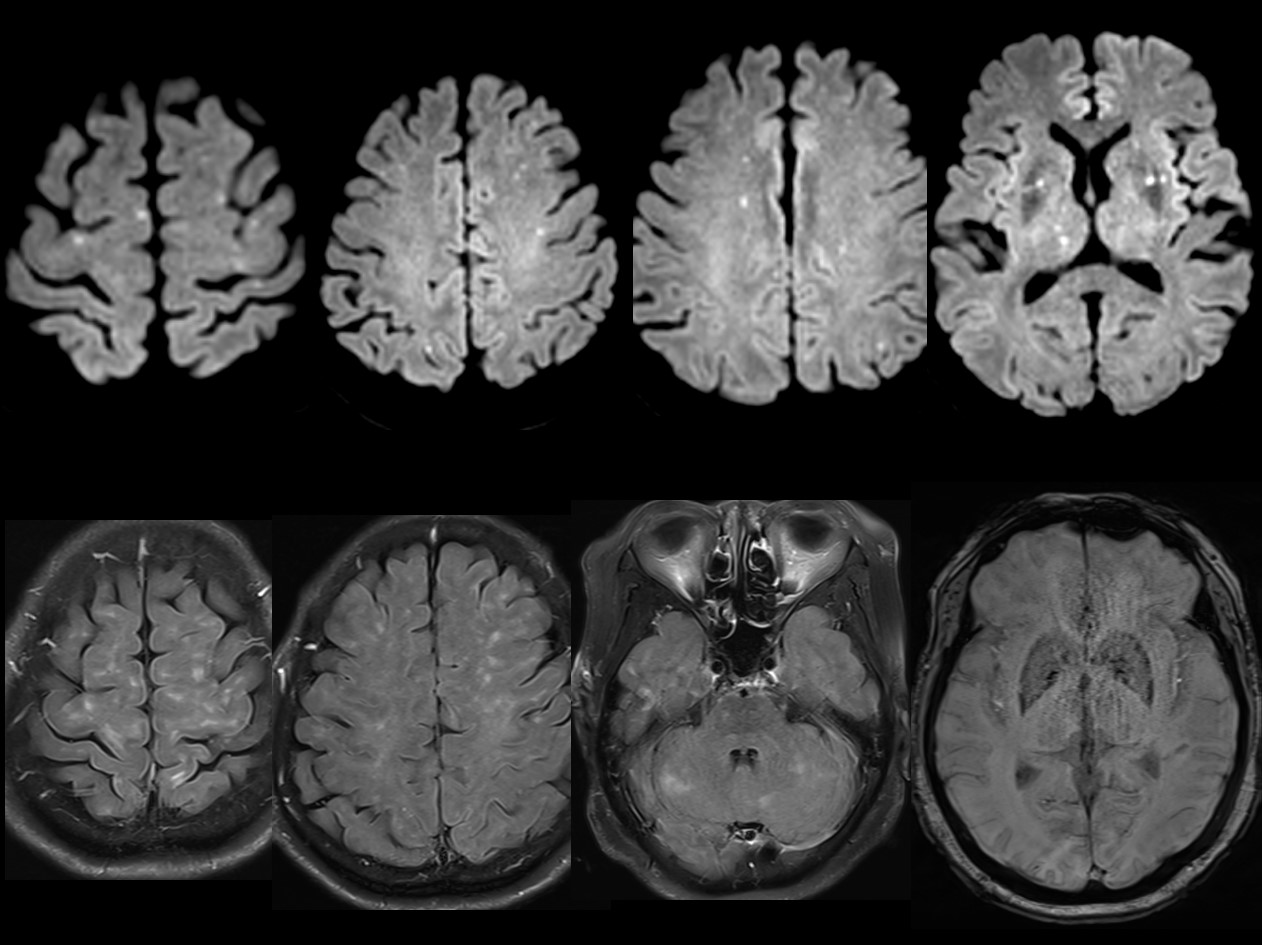
| Clinical information | 68세 여환 both TKA 3시간 후 시작된 drowsy mentality 로 시행한 Brain MRI이다. 진단은? |
| Discussion | Background: Fat embolism syndrome is a rare, life-threatening condition characterized by pulmonary, CNS, and skin symptoms. It occurs when fat obstructs arterial circulation, leading to delayed toxic injury from free fatty acids. After a long bone fracture, marrow fat enters the venous circulation, reaching the lungs and, with a right-to-left shunt, causing symptoms in the CNS, kidneys, eyes, and skin. Some cases of cerebral embolism occur without a shunt. Alternatively, inflammation may play a role, as lipoprotein lipase breaks down fat in the lungs, causing fat droplets to aggregate in microvessels, worsening organ dysfunction and potentially inducing cerebral cytotoxicity. Clinical Presentation: Neurologic symptoms in cerebral fat embolism are widely variable, ranging from headache, confusion, and seizure to coma Key Diagnostic Feature: Diagnosing cerebral fat embolism relies mainly on clinical evaluation, with neuroimaging playing a crucial role. Brain CT scans often appear normal, while MRI is the primary diagnostic tool. MRI analysis reveals evolving patterns, categorized into two major types. Type 1 (scattered cytotoxic edema) is the most common, with spot lesions showing restricted diffusion bilaterally in watershed areas and deep gray matter. Type 2 has three subtypes: 2A (confluent cytotoxic edema in white matter), 2B (vasogenic edema with hyperintensity), and 2C (petechial hemorrhage). Type 3 represents chronic sequelae, marked by brain parenchyma atrophy and hyperintense T2WI lesions without restricted diffusion, possibly indicating infarction, cavitation, scar formation, gliosis, or chronic demyelination. |
| Correct answer |
Correct Answer
Semi-Correct Answer |