HOME > Weekly Case > Weekly Case
Weekly Case
종결
Case No. 664
2023-08-01~2023-08-31
출제자 : 황인평
Hit : 493

| Period | 2023-08-01~2023-08-31 |
|---|---|
| Diagnosis | hemangioblastom |
| Gender |
|
| Age | 72 |
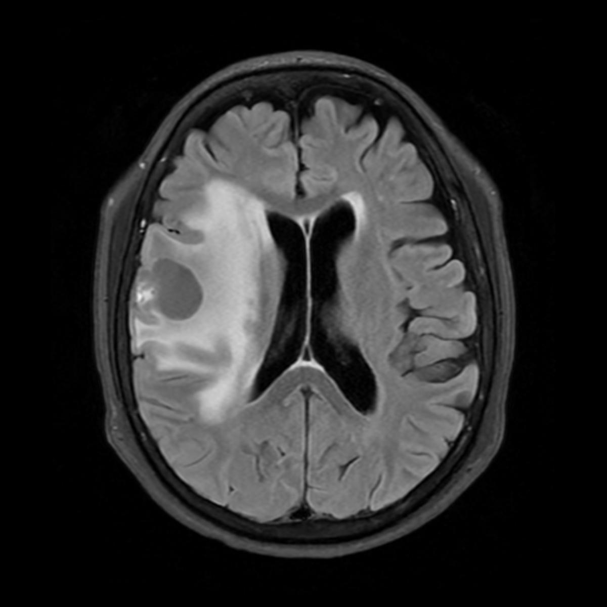
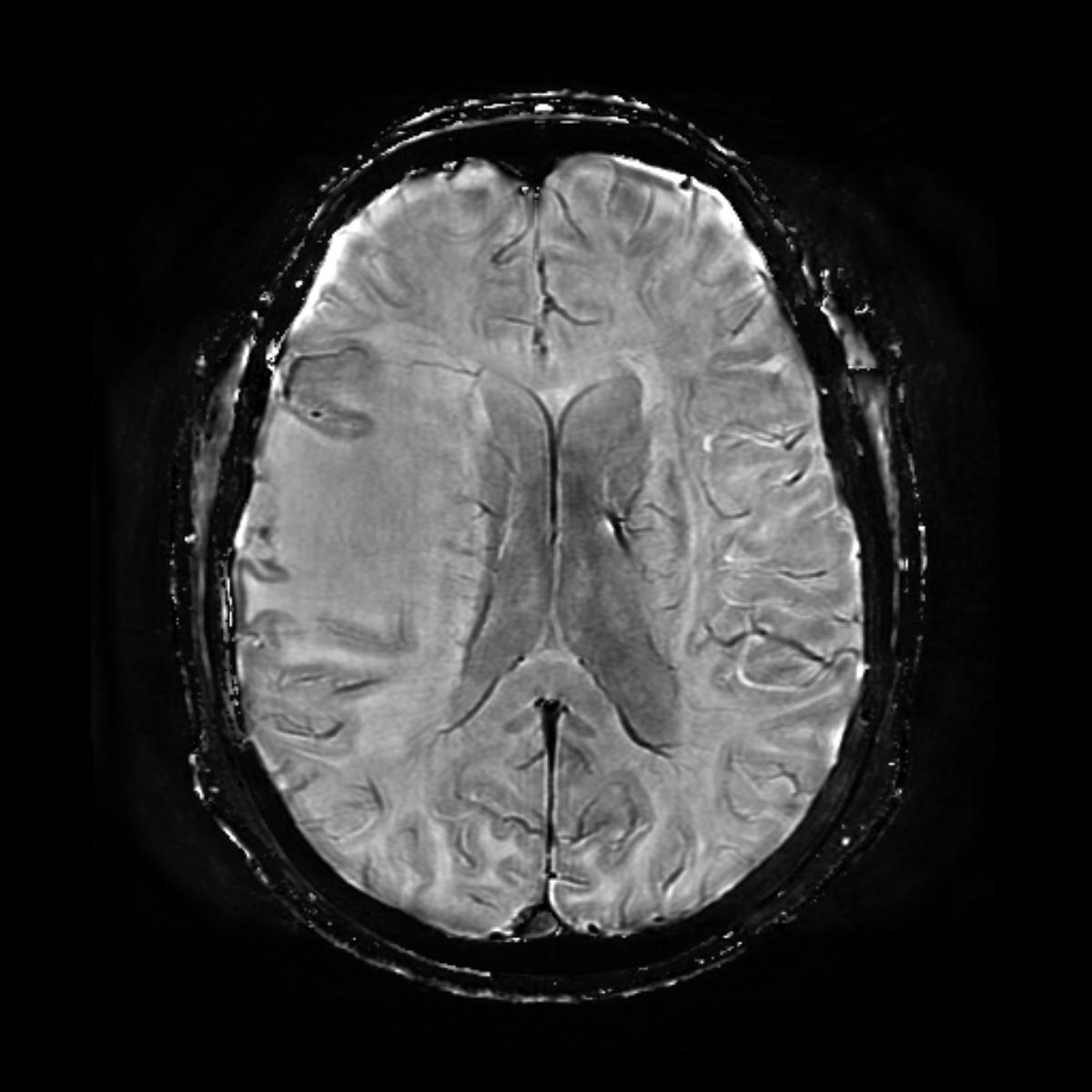
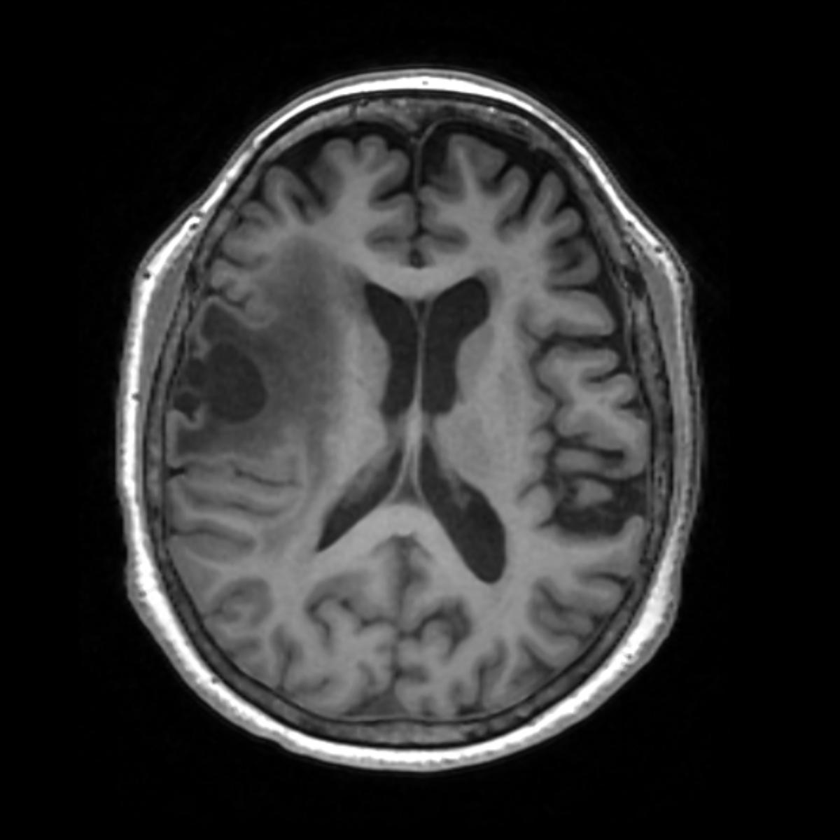
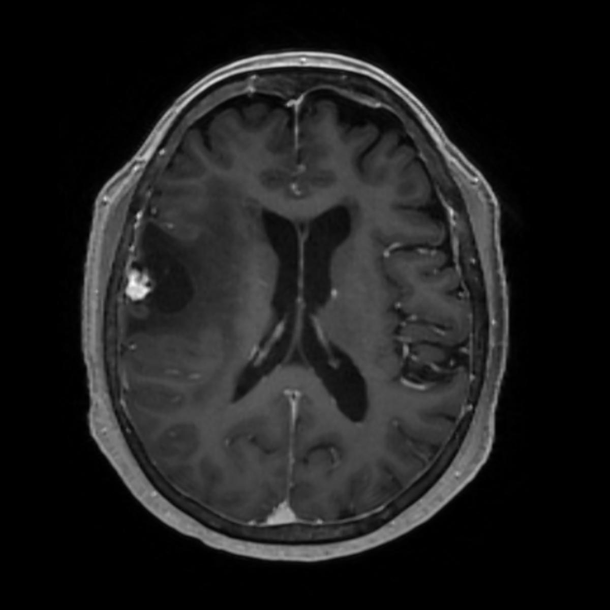
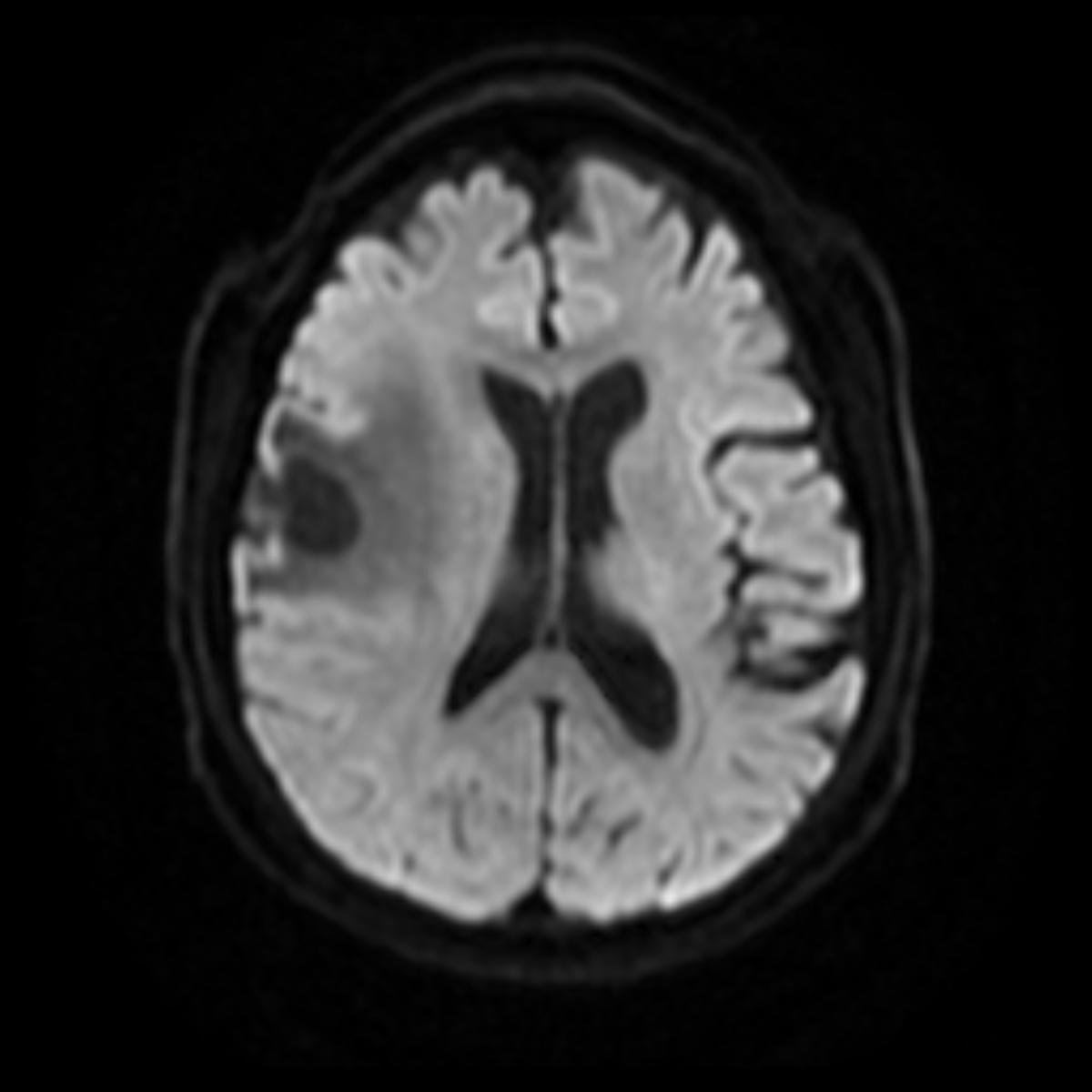
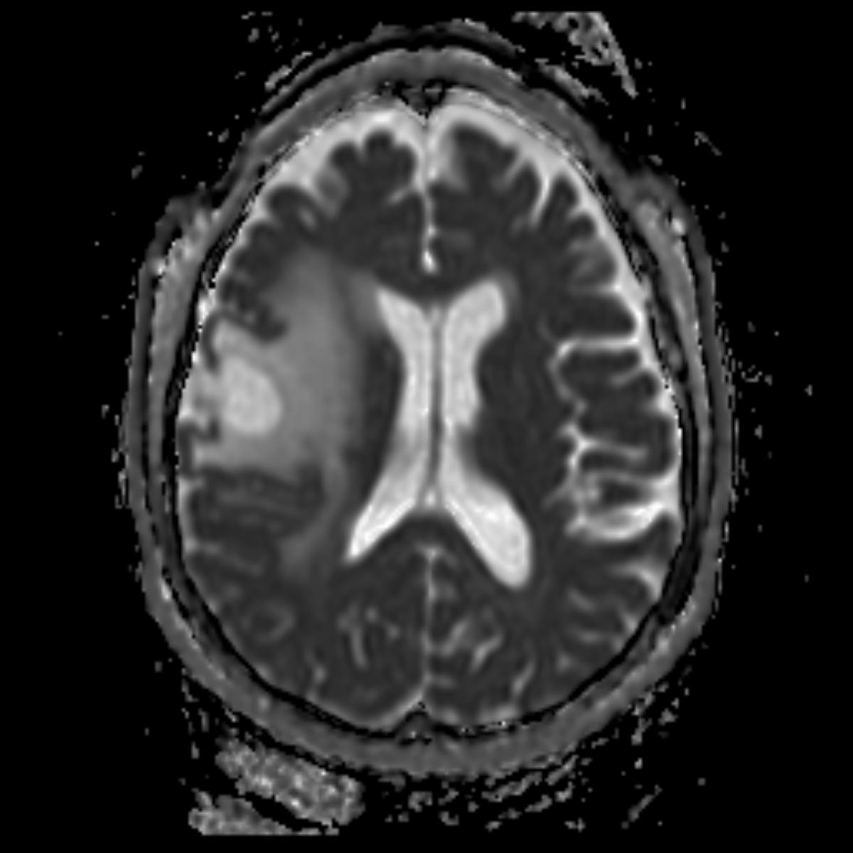
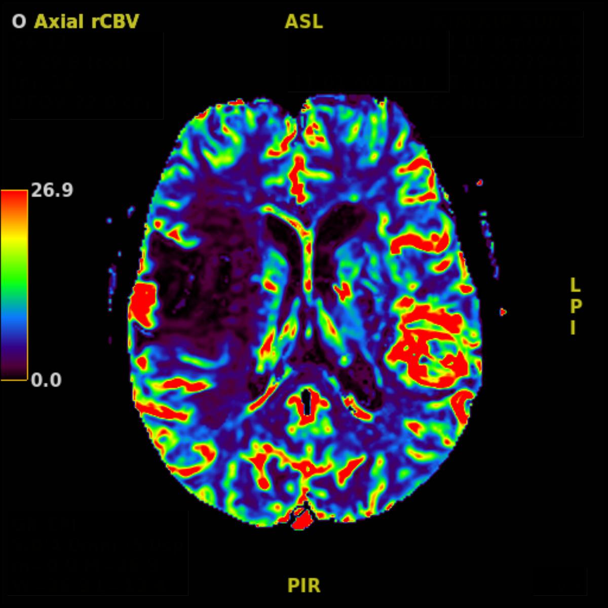
| Clinical information | F/72 C.C.: Lt. side central type facial palsy (onset: 10 hours ago) 진단은? |
| Discussion | Benign highly vascular intra-axial tumor. Most common posterior fossa primary tumor in middle aged/older adult. Location: Posterior fossa (90%-95%) > Supratentorial (5%-10%) Cerebellar hemisphere (80%) > vermis (15%) > medullar, 4th ventricle (5%) 25%-40% is associated with von Hipple-Lindau disease - Multiple hemangioblastoma (retinal and cerebral) - Renal cell carcinoma, pancreatic cysts and tumors Imaging finding: usually present as well-circumscribed masses with smooth margins - extratumoral cystic (a main cyst with a mural nodule) 63.3% - entirely solid 21.1% - intratumoral cystic 11.7% - mixed extratumoral and intratumoral cysts 3.9% MRI: - T1CE: strongly enhancing mural nodule - T2WI: hyperintense, flow voids due to enlarged vessels may be evident, especially at periphery of the cyst (60%-70%) - DWI: low signal / ADC: increased value - Angiography: extensive hypervascular nature Ref) J Korean Soc Radiol. 2016;75(1):26-29 Radiol Case Rep. 2022;17(11):4156–4160 |
| Correct answer |
Correct Answer
Semi-Correct Answer |