HOME > Weekly Case > Weekly Case
Weekly Case
종결
Case No. 653
2023-04-01~2023-04-30
출제자 : 임영희
Hit : 503
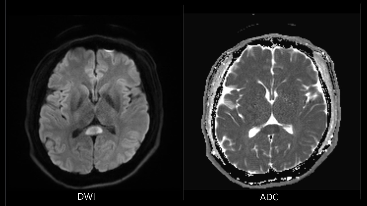
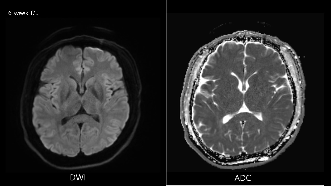
| Period | 2023-04-01~2023-04-30 |
|---|---|
| Diagnosis | Cytotoxic lesion of the corpus callosum |
| Gender |
|
| Age | 37 |
| Clinical information | 37/M Lt. sided hypesthesia 진단은? |
| Discussion | Clinical presentation Relates to the underlying pathology including seizure, metabolic disturbance, infection, CNS malignancy, drugs or toxins, results in cytotoxic edema. Imaging Transient splenium lesions, Well-circumscribed, small, oval lesions in midline within splenium. If more extensive, irregular lesions extending to adjacent hemisphere or anterior into body of corpus callosum. MRI T1: hypointense T2: hyperintense Diffusion restriction, no enhancement. Reference Starkey J, Kobayashi N, Numaguchi Y, Moritani T. Cytotoxic Lesions of the Corpus Callosum That Show Restricted Diffusion: Mechanisms, Causes, and Manifestations. Radiographics. 2017 Mar-Apr;37(2):562-576. doi: 10.1148/rg.2017160085. Epub 2017 Feb 6. PMID: 28165876. |
| Correct answer |
Correct Answer
Semi-Correct Answer |