HOME > Weekly Case > Weekly Case
Weekly Case
종결
Case No. 652
2023-04-01~2023-04-30
출제자 : 임영희
Hit : 431
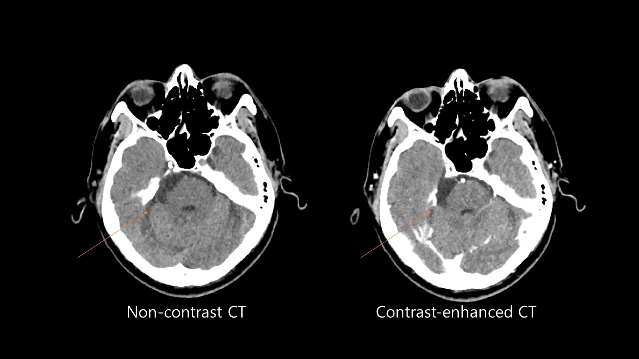
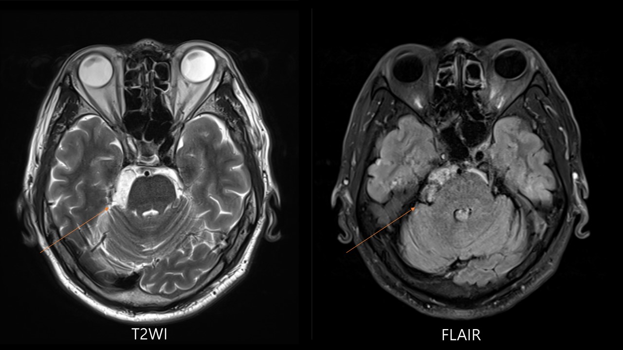
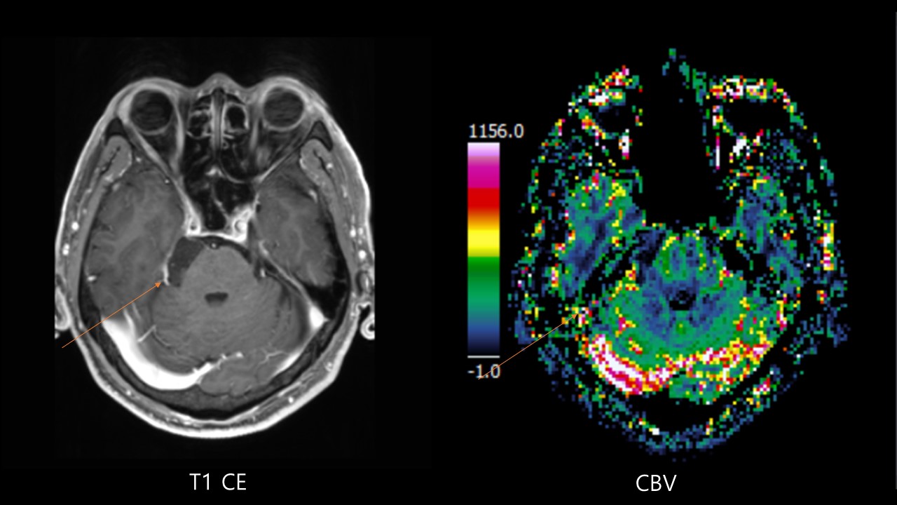
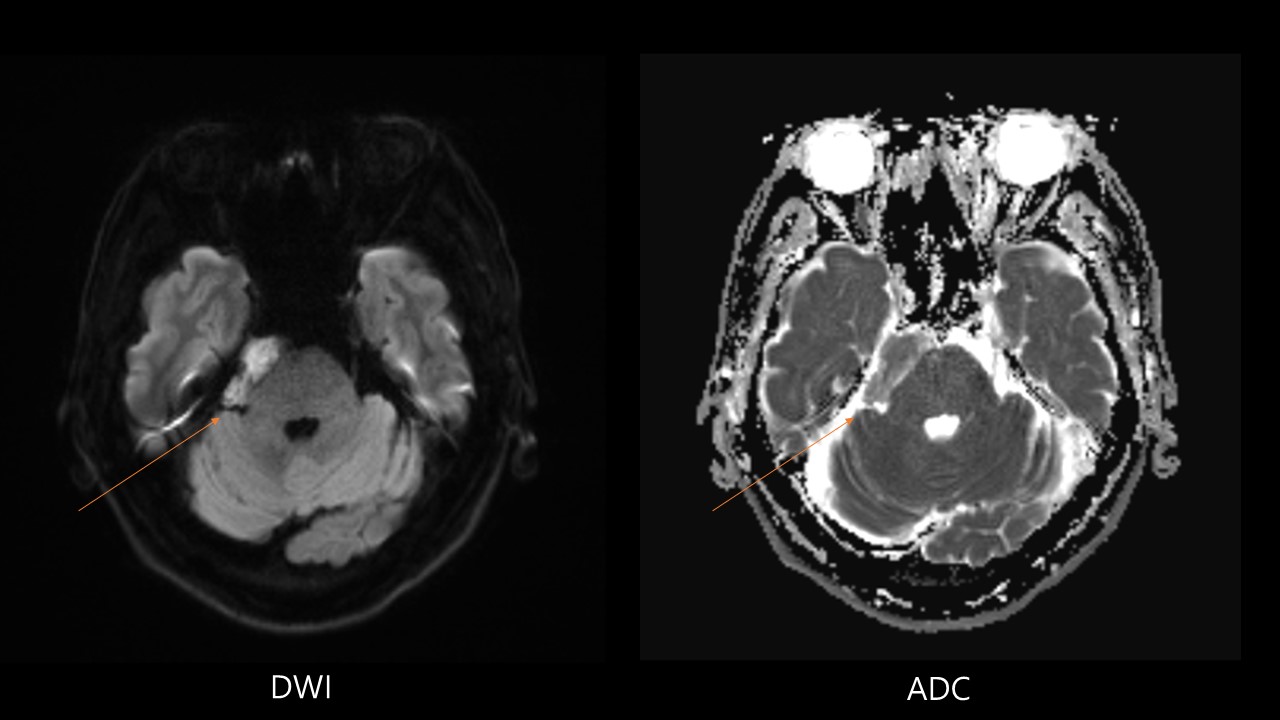
| Period | 2023-04-01~2023-04-30 |
|---|---|
| Diagnosis | epidermoid cyst |
| Clinical information | 51/M 우측 청력 저하, 5개월 전 진단은? |
| Discussion | CSF-like mass, insinuates cisterns and encases neurovascular structures. Lobulated, irregular, cauliflower-like mass Intradural (90%), primarily in basal cisterns Extradural (10%), skull and spine. 1. CT Round/lobulated mass > 95% hypodense, resembling CSF 10-25% contain calcifications Rare variant = “dense” epidermoid Usually non-enhancing, margin of cyst may show minimal enhancement. May have bony erosion 2. MR 2-1. T1WI Often (~ 75%) slightly hyperintense to CSF Uncommonly hyperintense to brain (“white epidermoid”) due to high triglycerides and unsaturated fatty acids Uncommonly hypointense to CSF (“black epidermoid”) ; Presence of solid crystal cholesterol and keratin, Lack of triglycerides and unsaturated fatty acids 2-2. T2WI Often isointense (65%) to slightly hyperintense (35%) to CSF Very rarely hypointense due to calcification, ↓ hydration, viscous secretions, and iron pigments 2-3. FLAIR; Usually does not completely null 2-4. DWI; Characteristic hyperintensity, ADC = brain parenchyma 2-5. T1WI C+ Usually none, though margin of cyst may show minimal enhancement (25%) Enhancing tumor is sign of malignant degeneration 2-6. MRS: Resonances from lactate, No NAA, choline, or lipid Reference Diagnostic Imaging: Brain, 3rd Edition Anne G Osborn & Miral D Jhaveri & Karen L. Salzman & A. James Barkovich |
| Correct answer |
Correct Answer
Semi-Correct Answer |