HOME > Weekly Case > Weekly Case
Weekly Case
종결
Case No. 646
2023-02-01~2023-02-28
출제자 : 유성혜
Hit : 483
| Period | 2023-02-01~2023-02-28 |
|---|---|
| Diagnosis | Desmoid-type fibromatosis in the head and neck |
| Gender |
|
| Age | 33 |
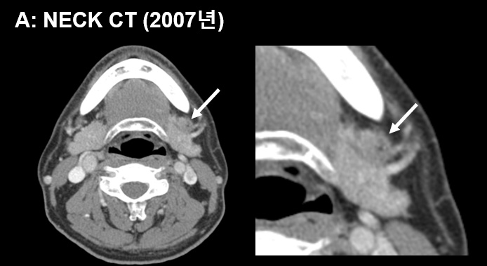
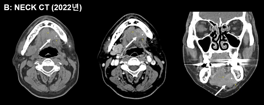
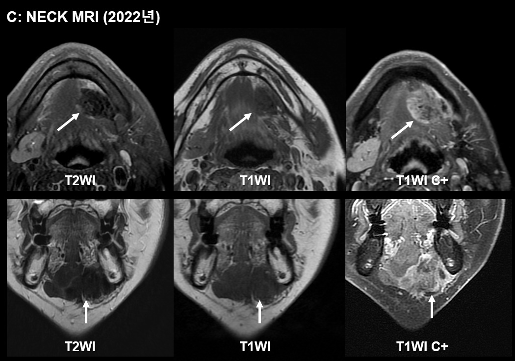
| Clinical information | Male / 33 문제: 2007년 그림 A에 보이는 mass에 대해 완전절제술을 받았던 환자로 2022년 좌측 턱밑 종물을 주소로 내원하여 CT 및 MRI를 시행하였다(그림 B, C). 진단은? |
| Discussion | Desmoid-type fibromatosis in the head and neck Benign fibroblastic tumor in the musculoaponeurotic system of the deep tissue. Although it is histopathologically benign and never metastasizes, the tumor is notorious for its infiltrative growth and local recurrence. MRI: T2WI: often high SI T1WI: iso SI T1WI C+: avid enhancement However, the MR signal intensity may be variable according to the degree of cellularity, myxoid change, and collagenization. Low signal intensity on T2-weighted image correlating with less cellularity and myxoid change with higher collagenization Reference) Rhim Jung hyo, Kim Ji-hoon, et al. Neuroradiology 2013;55:351-359 |
| Correct answer |
Correct Answer
Semi-Correct Answer |