HOME > Weekly Case > Weekly Case
Weekly Case
종결
Case No. 638
2022-10-01~2022-10-31
출제자 : 조세진
Hit : 586
| Period | 2022-10-01~2022-10-31 |
|---|---|
| Diagnosis | Solitary fibrous tumor |
| Gender |
|
| Age | 43 |
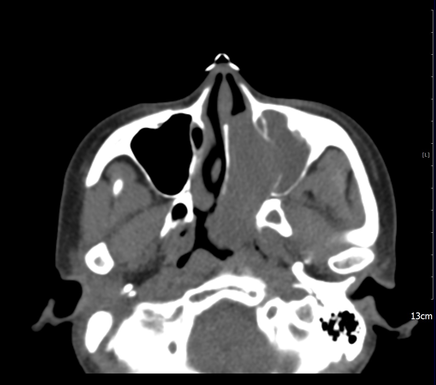
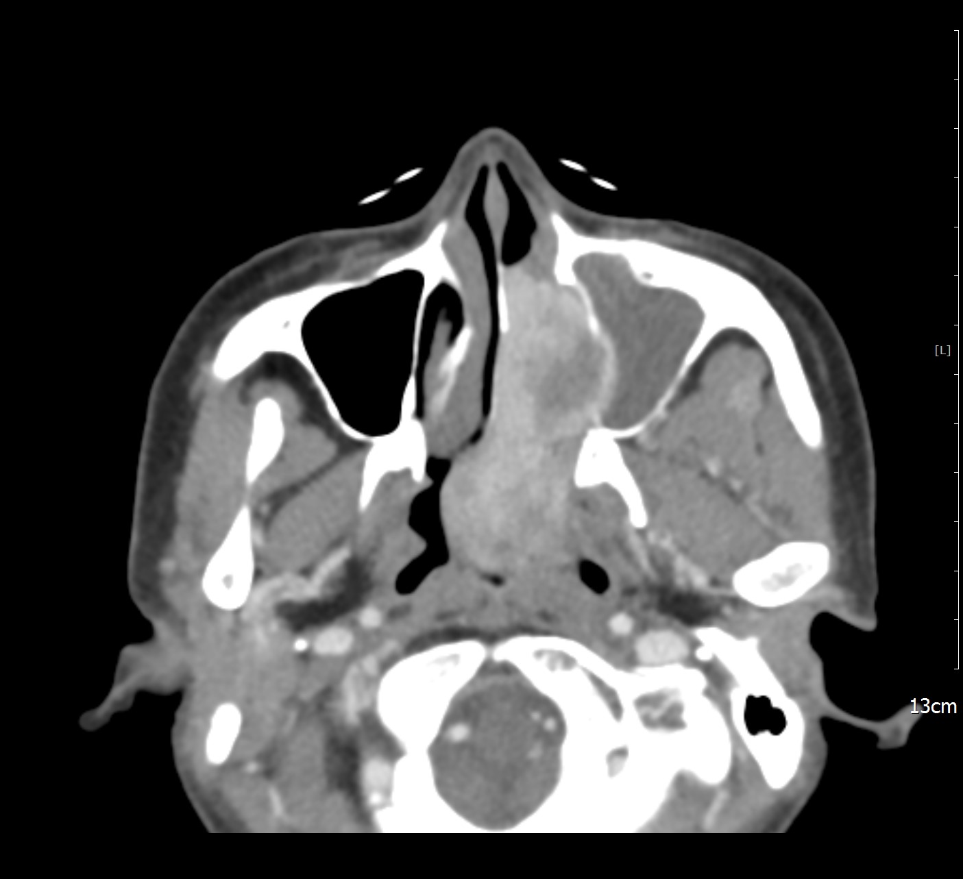
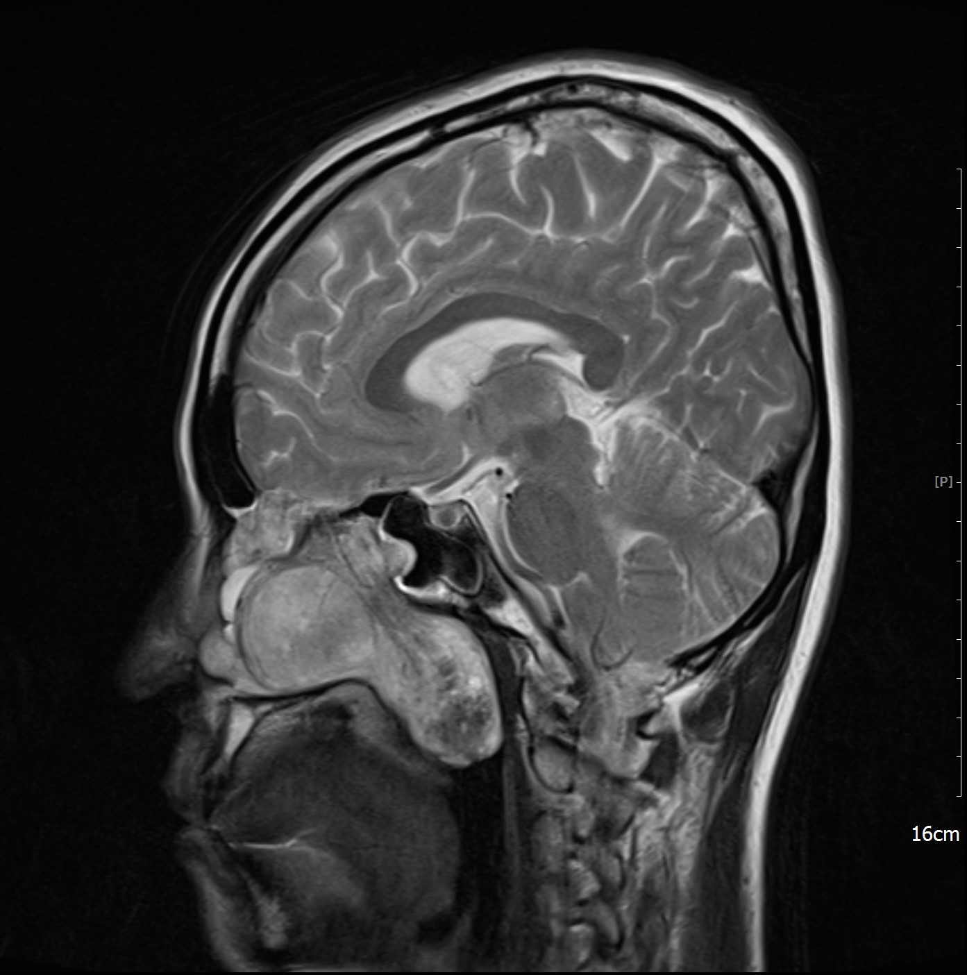
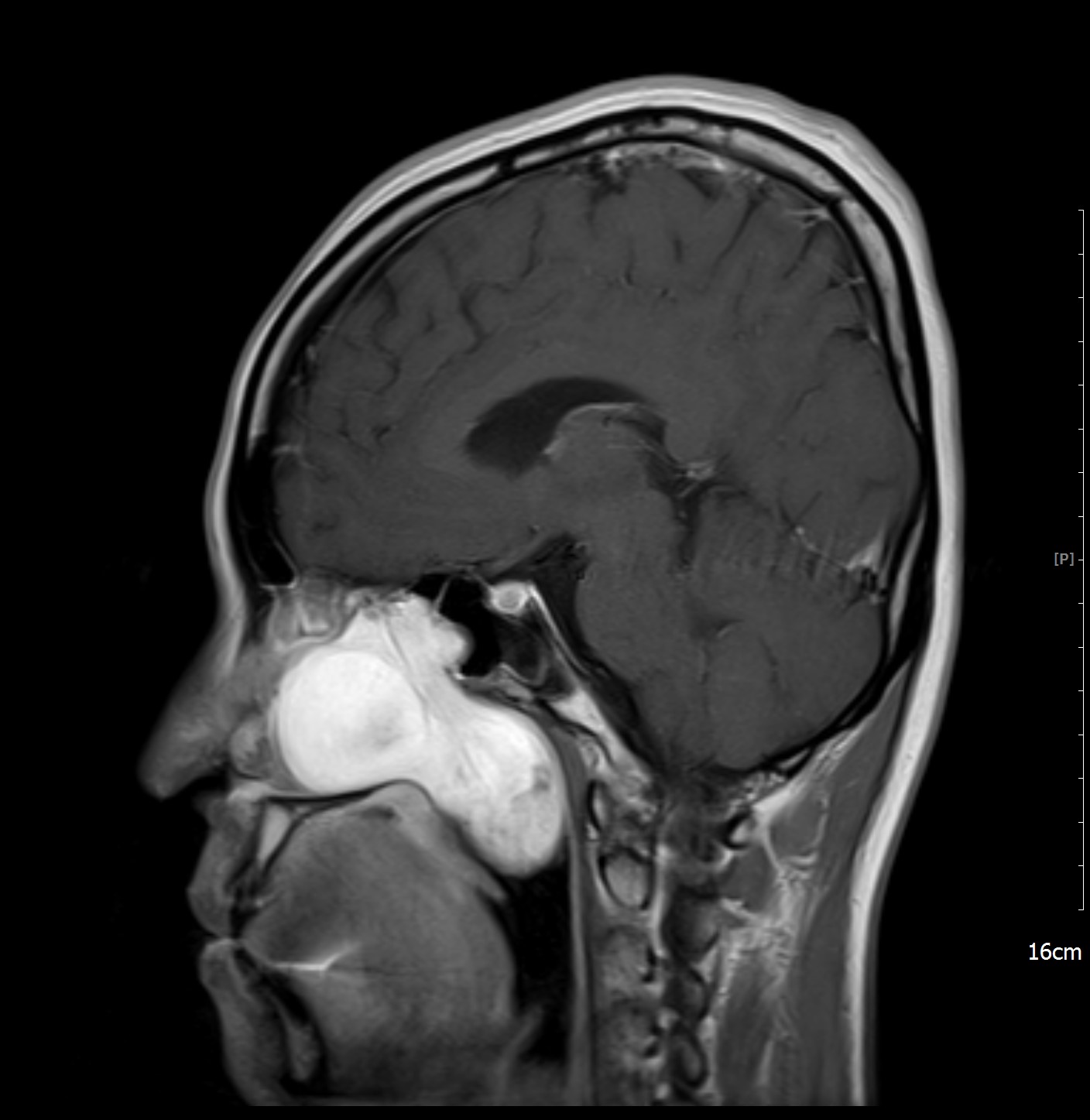
| Clinical information | 43/ F 임상정보: nasal cavity mass 진단은? |
| Discussion | Solitary fibrous tumor Review Uncommon spindle cell tumor of mesenchymal origin that usually arises in the pleura but can also arise in the head and neck Histopathologically, the lesions displayed plump spindle cells haphazardly intermixed in an attenuated collagenous stroma and prominent vascularity that resulted in a hemangiopericytoma-like pattern. Image findings - lobulated or well-defined margins - isodense to gray matter, may calcified, with bony erosion on CT - homogeneous signal intensity on pre-contrast T1WI, heterogeneous T2WI - may with intratumoral cystic areas - heterogeneous marked enhancement on Gd_T1WI - may show multiple tubular flow voids Ref) AJNR. June 2013, 34 (6) 1248-1251 |
| Correct answer |
Correct Answer
Semi-Correct Answer |