HOME > Weekly Case > Weekly Case
Weekly Case
종결
Case No. 631
2022-08-01~2022-08-31
출제자 : 황인평
Hit : 482
| Period | 2022-08-01~2022-08-31 |
|---|---|
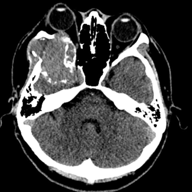
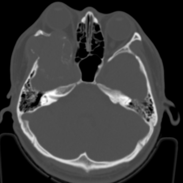
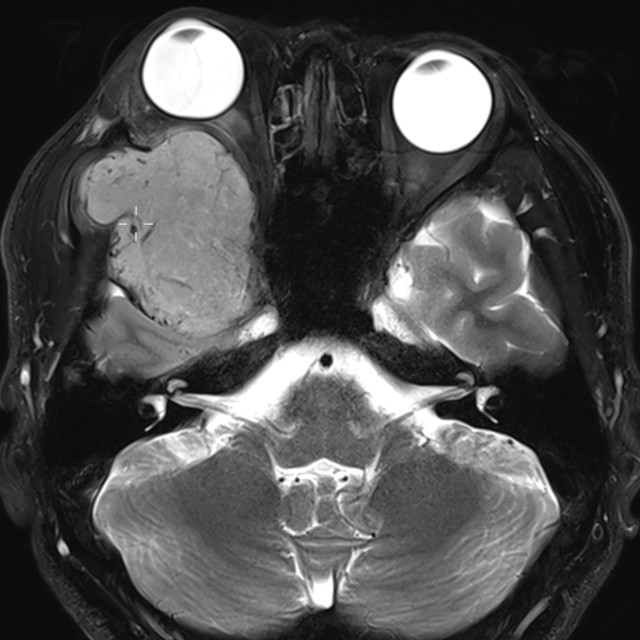
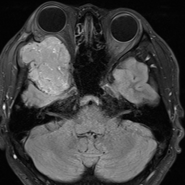
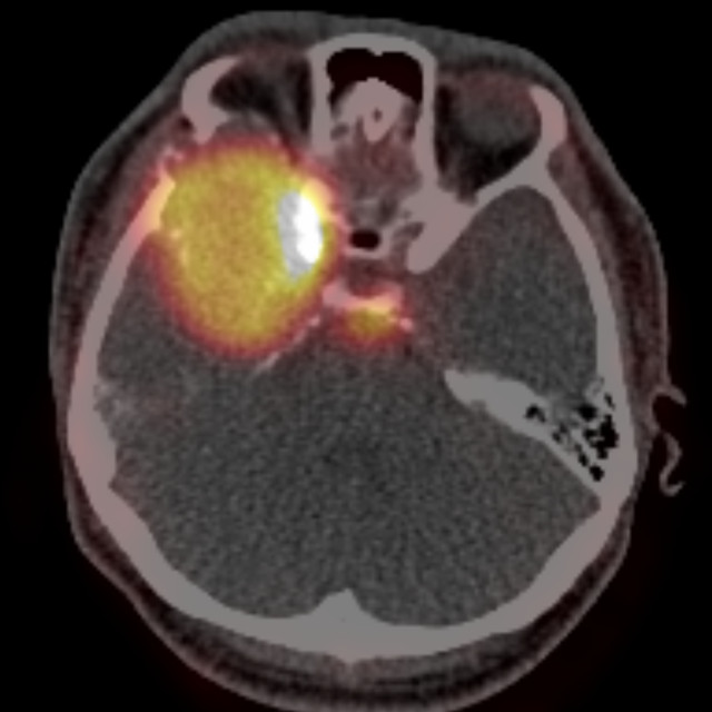
| Diagnosis | Intraosseous meningioma |
| Gender |
|
| Age | 25 |
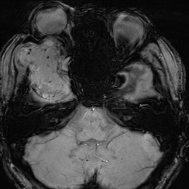
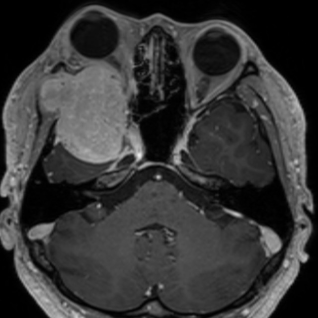
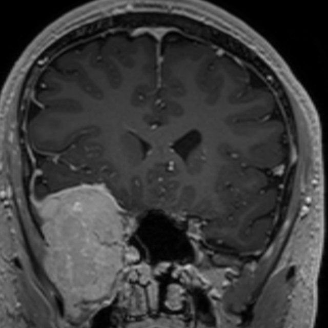
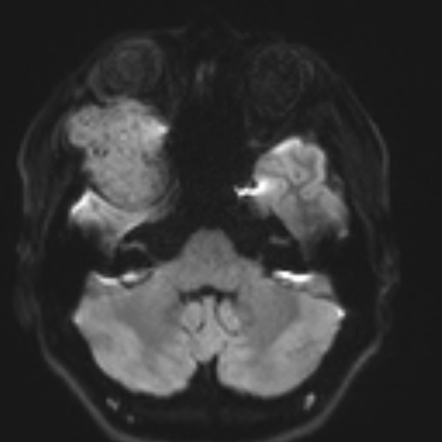
| Clinical information | M/25 C.C.: Right orbital pain (1MA) 진단은? |
| Discussion | Rare subtype of meningioma, most common type of extradural meningioma Common site : Frontoparietal, orbital regions Imaging Findings CT: Osteoblastic (~59%), osteolytic (~35%), or mixed type (~6%) Hyperostosis, atypical vascular marking, irregular foci of calcification MRI - T1: isointense extra-axial mass component with the expanded bony component being low signal similar to the rest of the skull - T2: isointense to grey matter while a small proportion can be hyperintense - T1 C+ (Gd): uniform avid contrast enhancement, dural enhancement. Ddx. For osteolytic intraosseous meningioma metastatic cancer, plasmacytoma, giant cell tumor, hemangioma, epidermoid cyst, osteogenic sarcoma, eosinophilic granuloma, aneurysmal bone cyst, or fibrous dysplasia, intraosseous malignant meningioma. Reference. AJNR Am J Neuradiol. 2005;26(8): 2053-56 |
| Correct answer |
Correct Answer
Semi-Correct Answer |