HOME > Weekly Case > Weekly Case
Weekly Case
종결
Case No. 623
2022-04-01~2022-04-30
출제자 : 임영희
Hit : 944
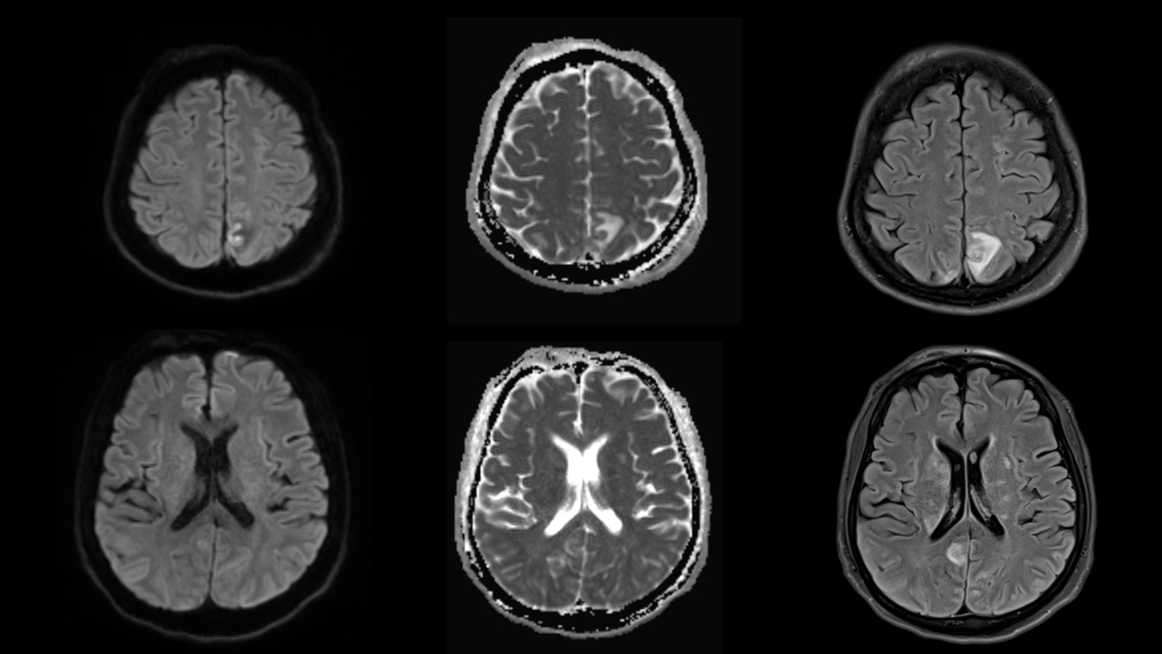
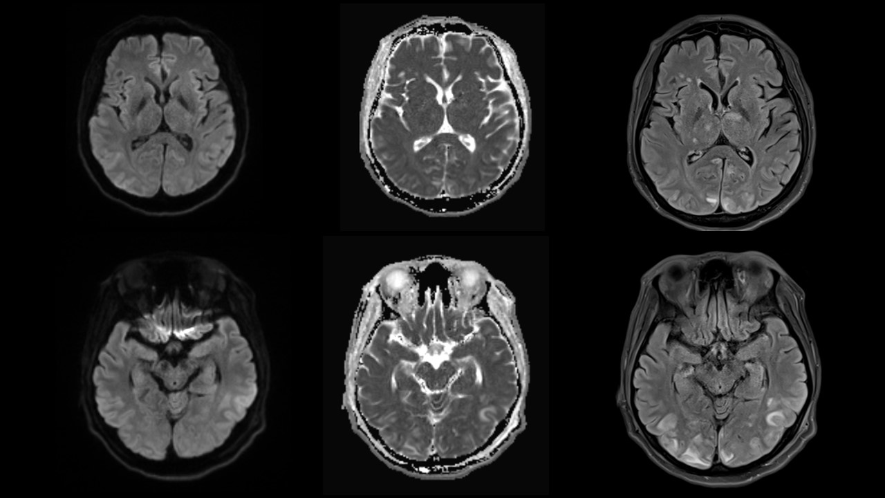
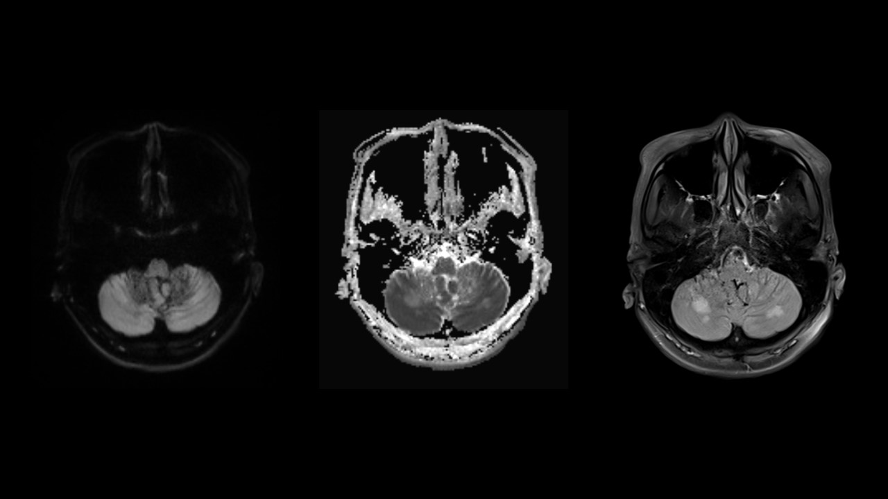
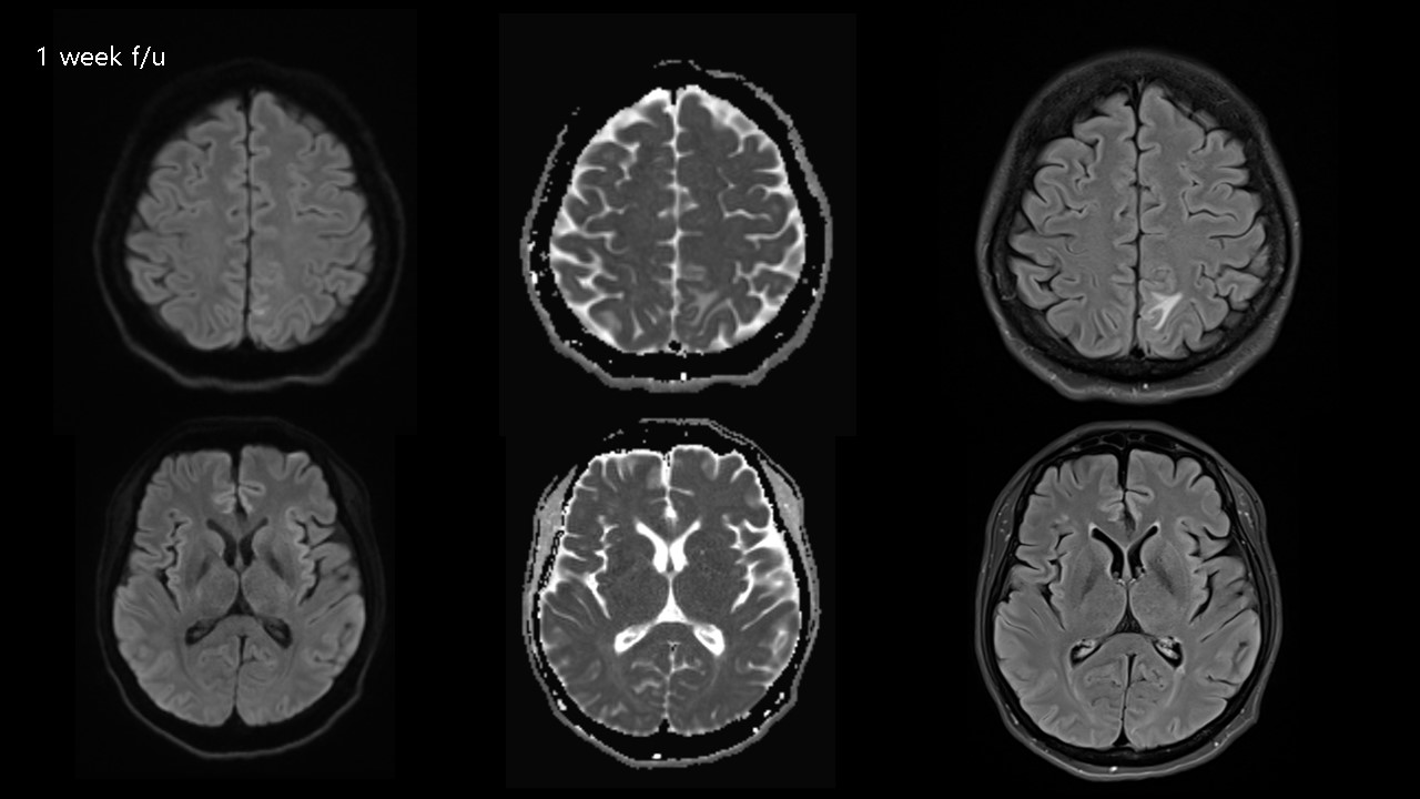
| Period | 2022-04-01~2022-04-30 |
|---|---|
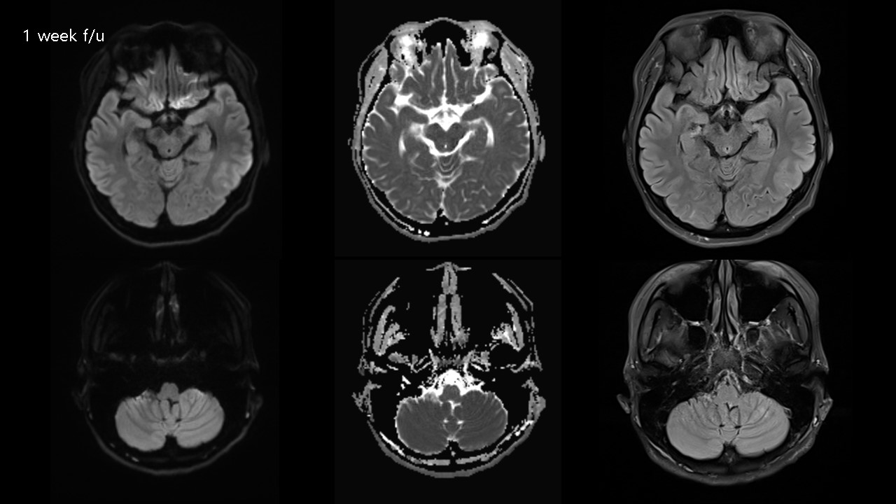
| Diagnosis | PRES (Posterior reversible encephalopathy syndrome) |
| Gender |
|
| Age | 37 |
| Clinical information | 37/F 의식저하 (history of end stage kidney disease) |
| Discussion | Definition Variant of hypertensive encephalopathy characterized by headache, visual disturbances, altered mental function Cerebrovascular autoregulatory disorder; Multiple etiologies, Most caused by acute hypertension (HTN) # Image findings -CT Bilateral nonconfluent hypodense foci, predilection to posterior parietal, occipital lobes and cortical watershed zones. -CTA Major vessels usually normal Distal vessels may show diffuse vasoconstriction, focal irregularity and beaded appearance. - MRI Typical PRES: T2/FLAIR Hyperintense cortical/subcortical lesions, Parietooccipital lobes, cortical watershed zones Atypical PRES: T2/FLAIR lesion in frontal lobe, basal ganglia involvement, Extensive brainstem, cerebellar edema “Leaky” blood-brain barrier may cause gadolinium accumulation in CSF, FLAIR hyperintensity T2* GRE 3 patterns of hemorrhage: Focal parenchymal hemorrhage, microhemorrhages, convexity subarachnoid hemorrhage (SAH) DWI Most common: No restriction Less common: Hyperintense on DWI with “pseudonormalized” ADC, May indicate irreversible infarction ADC map: Markedly elevated (bright areas) PWI; May show ↑ rCBV T1WI C+; Variable patchy enhancement MRS; May show widespread metabolic abnormalities, ↑ Cho, Cr, mildly ↓ NAA, Usually return to normal within 2 months Reference Diagnostic Imaging: Brain, 3rd Edition Anne G Osborn & Miral D Jhaveri & Karen L. Salzman & A. James Barkovich |
| Correct answer |
Correct Answer
Semi-Correct Answer |