HOME > Weekly Case > Weekly Case
Weekly Case
종결
Case No. 622
2022-04-01~2022-04-30
출제자 : 임영희
Hit : 652
| Period | 2022-04-01~2022-04-30 |
|---|---|
| Diagnosis | brain abscess |
| Clinical information | 70/M LOC, GTC seizure 진단은? |
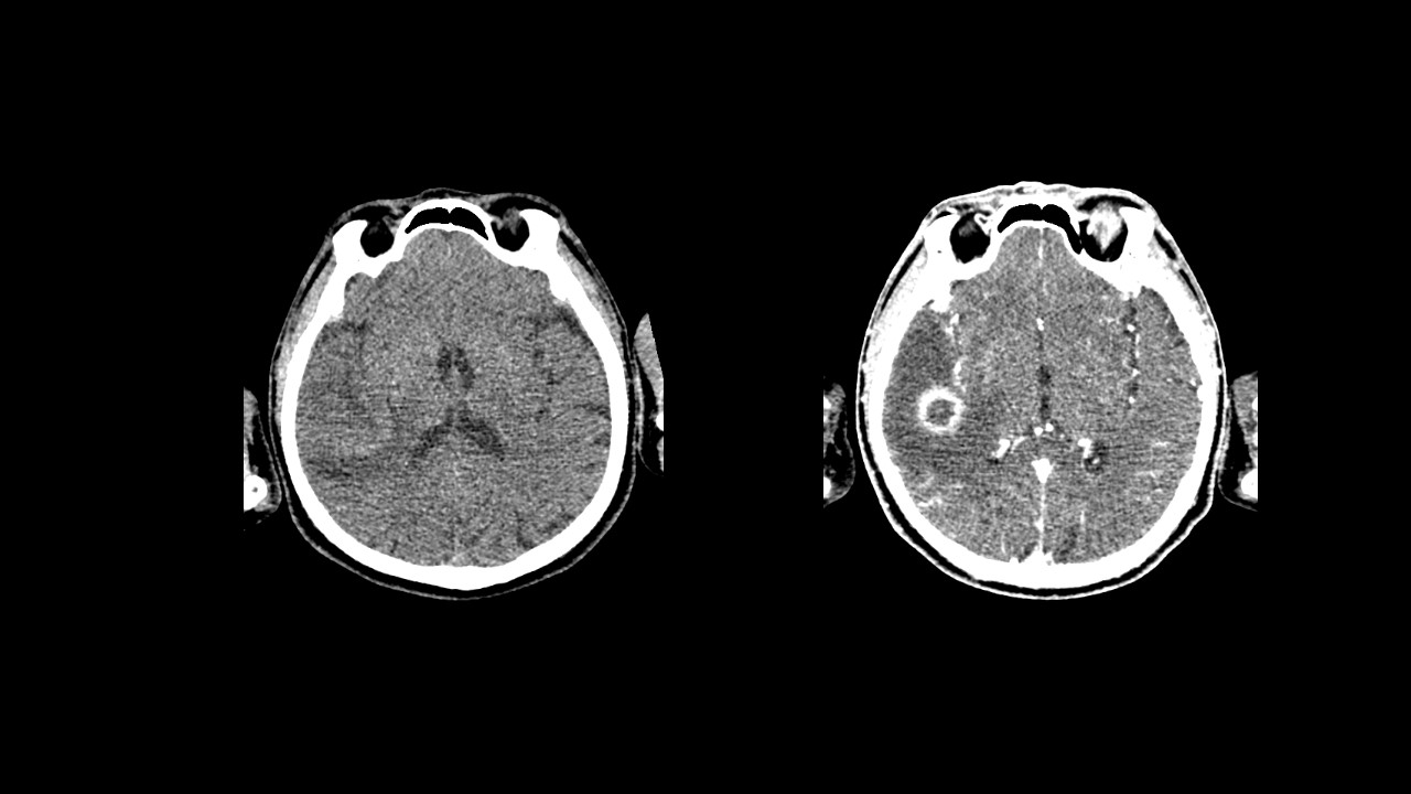
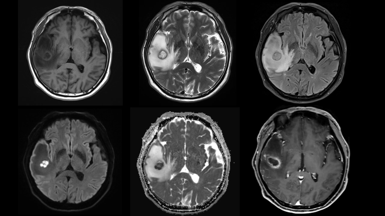
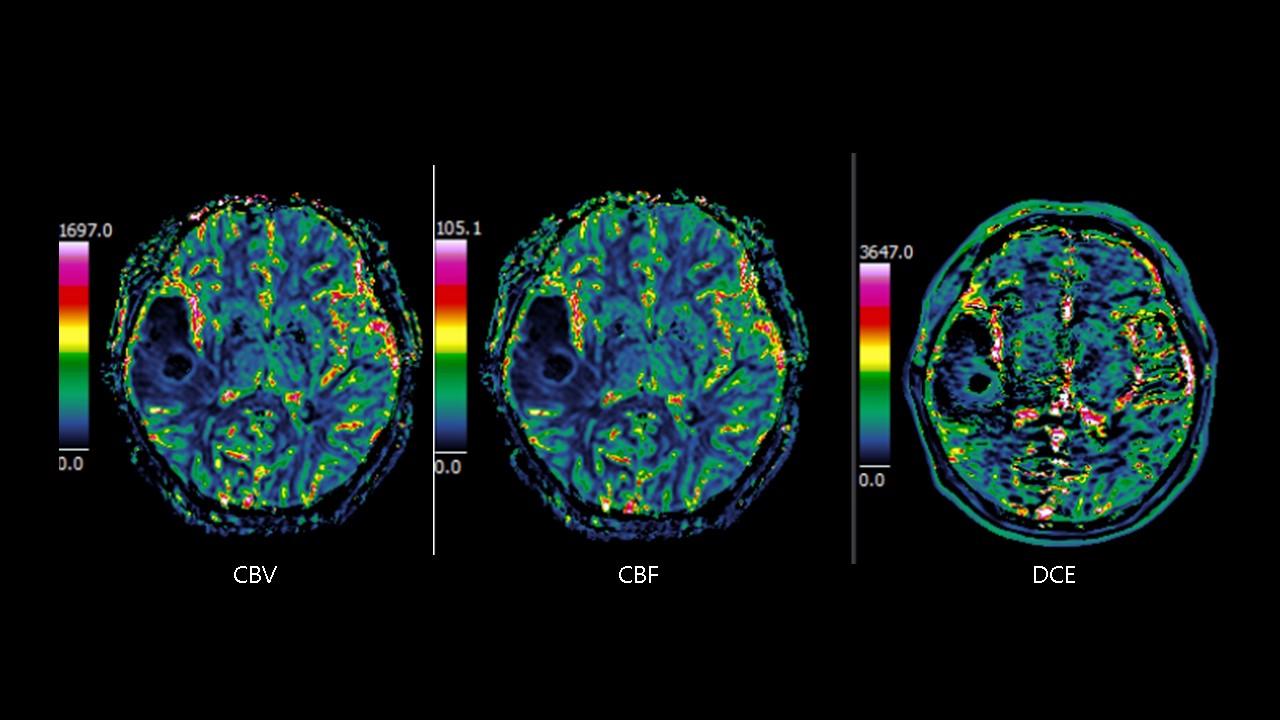
| Discussion | Definitions Localized infection of brain parenchyma, typically bacterial; fungal or parasitic less common 4 pathologic stages: Early cerebritis, late cerebritis, early capsule, late capsule # Image findings 1>Early cerebritis CT: Ill-defined, hypodense subcortical lesion with mass effect; CT may be normal early MR:Poor marginated T1 hypo-iso SI, T2 hyper SI, Patchy enhancement 2>Late cerebritis CT: Central low-density area; peripheral edema, increased mass effect, Irregular rim enhancement MRL T1 Hypointense center, iso-/mildly hyperintense rim, T2 Hyperintense center, hypointense rim; hyperintense edema, Intense, irregular rim enhancement. 3> Early capsule CT: Low-density center with thin, distinct enhancing rim MR: T1 Rim iso-/hyperintense to white matter (WM); center hyperintense to CSF T2 Hypointense rim (due to collagen, hemorrhage, or paramagnetic free radicals) Well-defined, thin-walled enhancing rim 4> Late capsule CT: shrinks, capsule thickens, MR: Cavity shrinks, capsule thickens DWI: Increased signal intensity in cerebritis and abscess ADC map: Markedly decreased signal (low ADC) centrally within abscess MRS; Central necrotic area may show presence of amino acids (0.9 ppm), lactate (1.3 ppm), acetate (1.9 ppm), succinate (2.4 ppm) PWI: Abscess capsule shows lower rCBV values than rims of enhancing tumors Reference Diagnostic Imaging: Brain, 3rd Edition Anne G Osborn & Miral D Jhaveri & Karen L. Salzman & A. James Barkovich |
| Correct answer |
Correct Answer
Semi-Correct Answer |