HOME > Weekly Case > Weekly Case
Weekly Case
종결
Case No. 621
2022-04-01~2022-04-30
출제자 : 임영희
Hit : 448
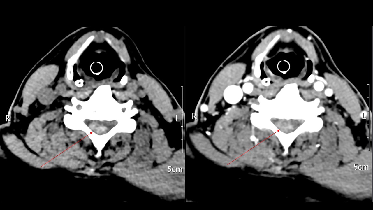
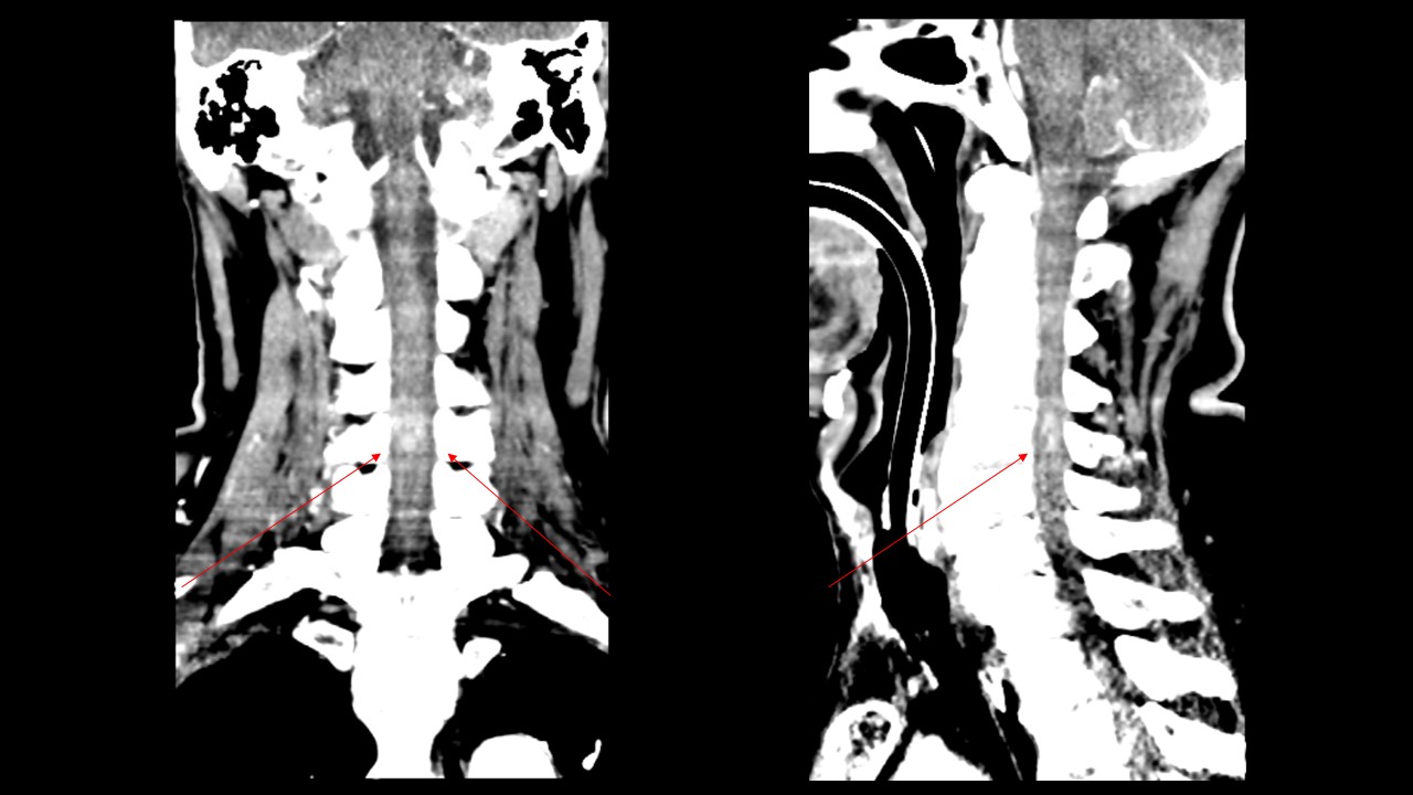
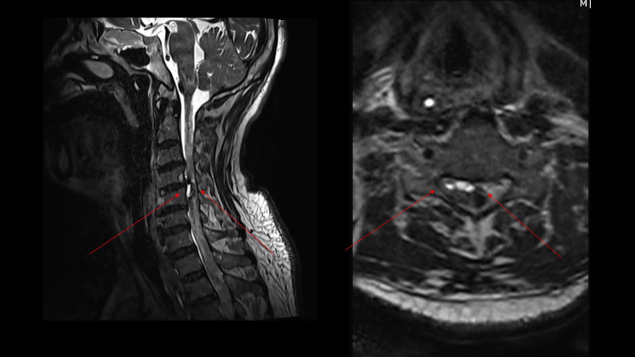
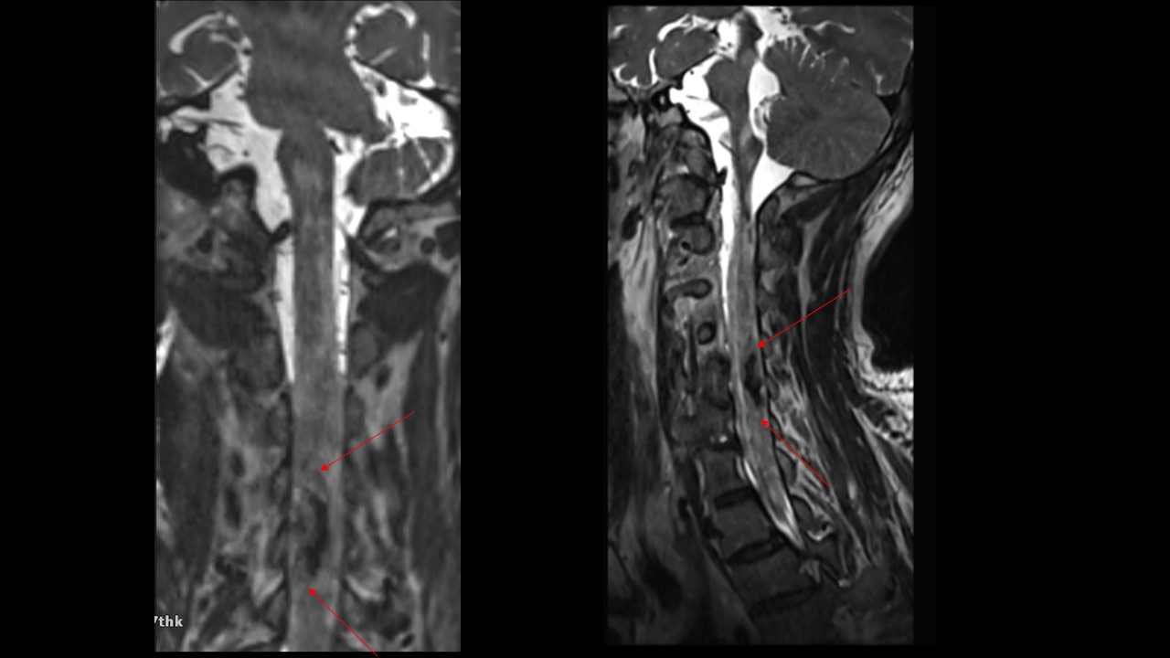
| Period | 2022-04-01~2022-04-30 |
|---|---|
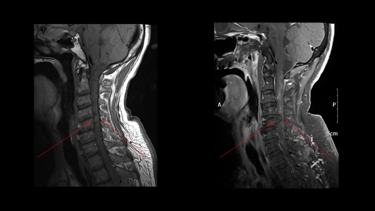
| Diagnosis | spinal cavernous malformation |
| Gender |
|
| Age | 64 |
| Clinical information | 64/M Upper extremity numbness 진단은? |
| Discussion | Vascular lesion with lobulated, thin sinusoidal vascular channels and no interspersed neural tissue Spinal cord uncommon site: 3-5% of all cavernous malformations, most located in thoracic spine Annual hemorrhage risk is 2.1%. Synonyms - Cavernous angioma, cavernous hemangioma, “cryptic” vascular malformation # Image findings Locules of blood with fluid0fluid levels surrounded by T2-hypointense rim. MR Conventional angiography – usually negative T1WI Heterogeneity due to blood products of varying ages Typical speckled “popcorn” heterogeneous signal Small lesion may not show obvious heterogeneous signal T2WI Heterogeneous, hypointense hemosiderin rim Small lesion may not show obvious heterogeneous signal, may show only focal low signal Typically no edema unless recent hemorrhage T2* GRE Prominent “blooming” due to susceptibility effects T1WI C+ Enhancement absent/minimal Reference Jeffrey S. Ross & Kevin R. Moore, Diagnostic Imaging: Spine, 3rd Edition |
| Correct answer |
Correct Answer
Semi-Correct Answer |