HOME > Weekly Case > Weekly Case
Weekly Case
종결
Case No. 615
2022-02-01~2022-02-28
출제자 : 조세진
Hit : 465
| Period | 2022-02-01~2022-02-28 |
|---|---|
| Diagnosis | Sinonasal Ewing sarcoma |
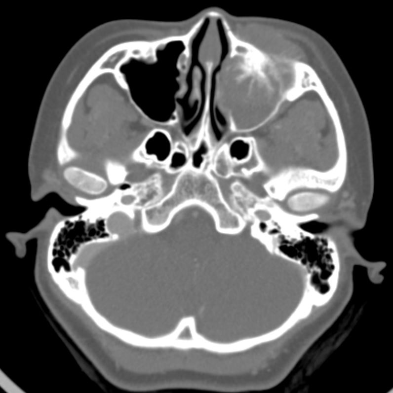
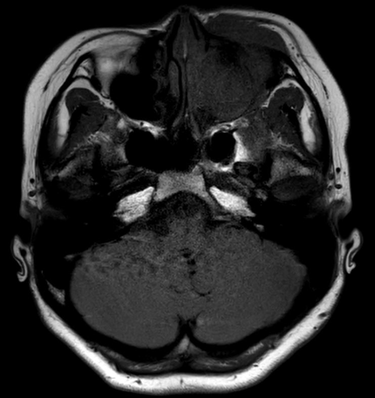
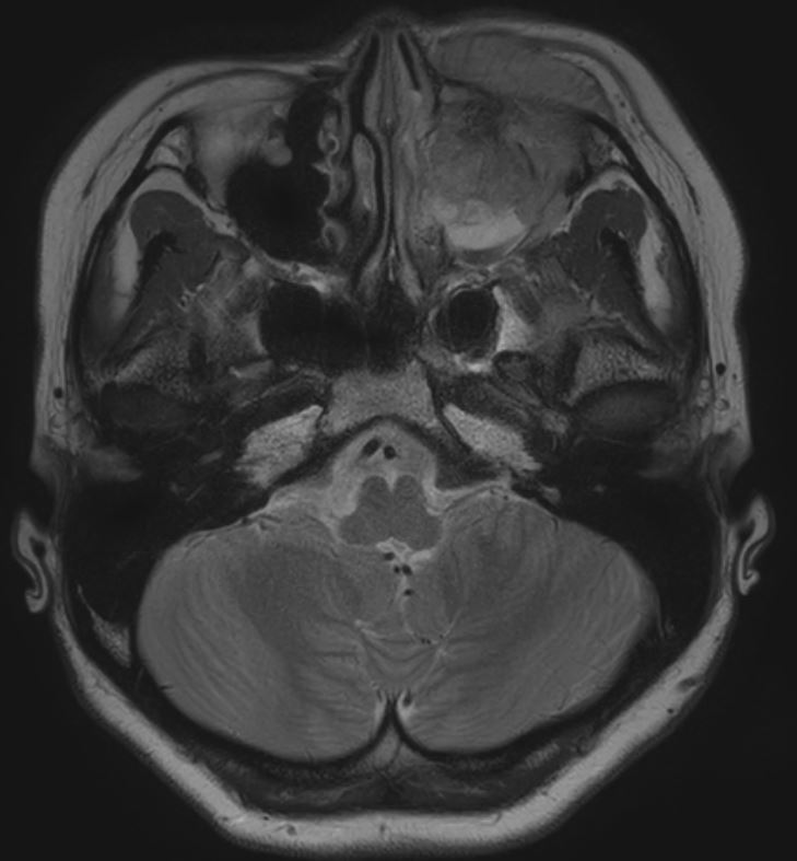
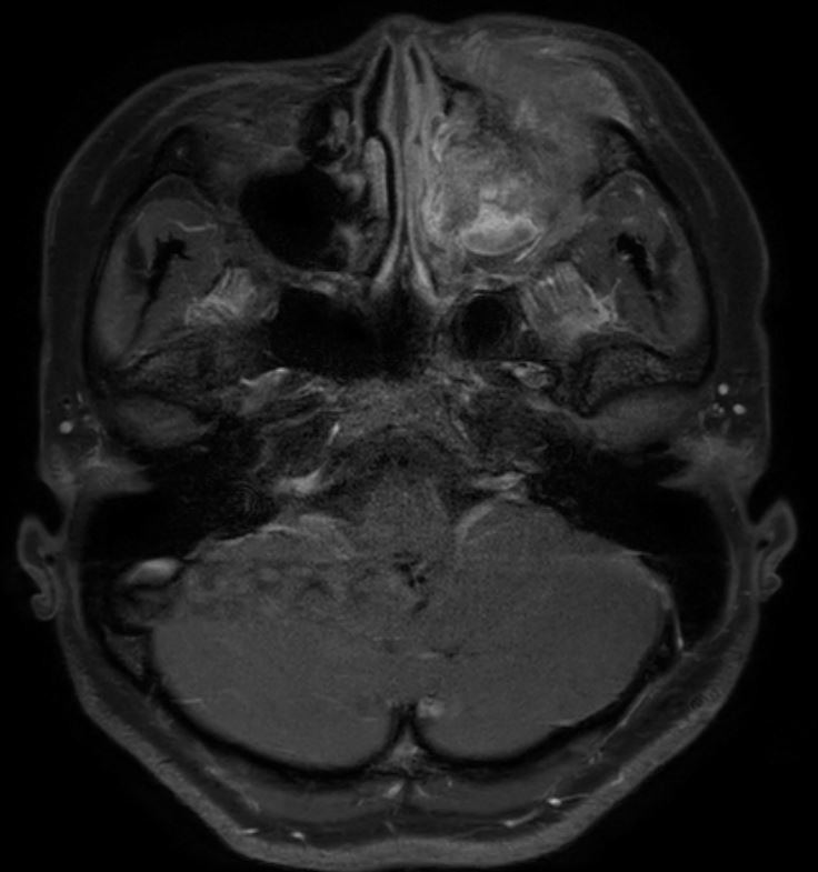
| Clinical information | Question> F/17Y Symptom: Facial pain, numbness 얼굴 통증을 주소로 내원한 17세 여자 환자의 영상이다. 진단은? Images> axial CT, axial T1WI, axial T2WI, axial Gd_T1WI |
| Discussion | Sinonasal Ewing sarcoma: - A rare malignant disease, making up only 4% to 6% of all primary bone tumors. - Involves the head and neck region in only 1% to 4% (mandible or maxilla (1%–2%), facial bones (0.5%) of ES cases, and tumors with a sinonasal origin form another rare subset. - Osseous lesions originate in the central medullary canal, with early involvement of the soft tissues in 80%–100% of cases. Soft-tissue masses are usually large and circumferential about the involved bone; they may exceed the intraosseous component in size in 2%–15% of cases. The communication between the medullary canal and soft-tissue components may be through focal cortical destruction or through permeation of the cortical haversian canal system and along neurovascular channels with small nests of tumor cells. Periosteal elevation is common. Imaging findings: Bone marrow replacement, cortical bone destruction, moth-eaten to permeative pattern, wide zone of transition (poor margination), periosteal reaction (either lamellated (onionskin) or spiculated (sunburst or hair-on-end)), associated soft-tissue mass formation area common. - T1: low to intermediate signal, T1 C+ (Gd): heterogeneous but prominent enhancement T2: heterogeneously high signal, may see hair on end low signal striations Treatment : Systemic chemotherapy is the mainstay of treatment |
| Correct answer |
Correct Answer
Semi-Correct Answer |