HOME > Weekly Case > Weekly Case
Weekly Case
종결
Case No. 612
2022-01-01~2022-01-31
출제자 : 서종현
Hit : 444
| Period | 2022-01-01~2022-01-31 |
|---|---|
| Diagnosis | Toxoplasmosis |
| Gender |
|
| Age | 27 |
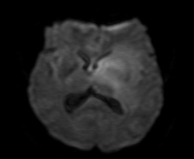
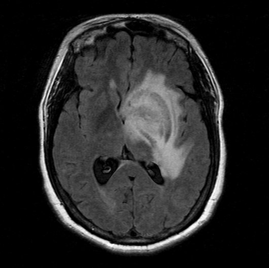
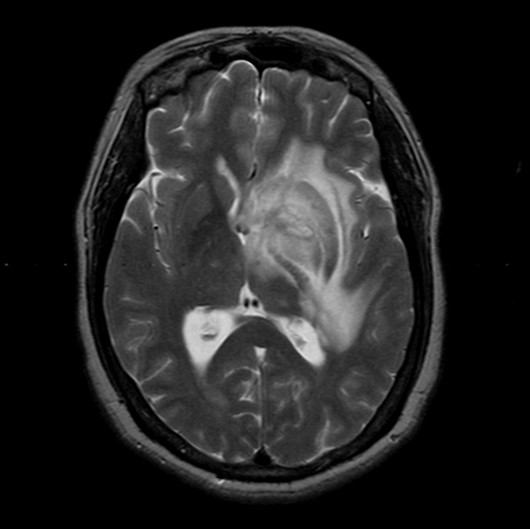
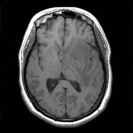
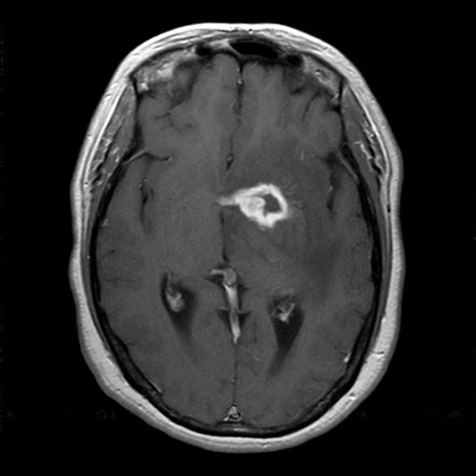
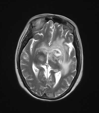
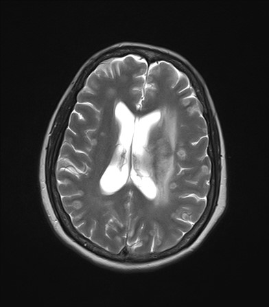
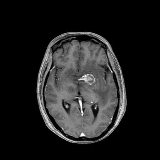
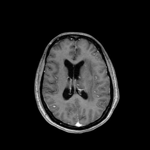
| Clinical information | C.C>균형을 못 잡고, 글 인지를 잘 못하는 27세 남자환자이다. 3개월전 HIV를 진단받았다. 진단은? (Fig 1~5=>initial, Fig6-9=>FU) |
| Discussion | Toxoplasmosis Brain biopsy에서 high grade glioma 의심소견이 나왔으나 이후 HIV가 진단되었고 F/U에서dissemination된 병변이 생김. CSF study에서 toxoplasma DNA PCR에서 positive 나와 CNS toxoplasmosis로 진단됨. 1. Introduction Caused by Toxoplasma gondii: ubiquitous intracellular parasite m/c opportunistic CNS infection in AIDS patients, immunocompromised patient - Nearly 1/4 of T. gondii-seropositive AIDS patients develop toxoplasmosis m/c location: basal ganglia, thalamus, corticomedullary junction, periventricular region Histologic findings: poorly circumscribed necrotizing, coagulative abscess with hyperemic border Treatment: pyrimethamine + sulfadiazine (resolution in 2-4 weeks) 2. Radiologic finding T1: low SI, mild peripheral SI (coagulative necrosis or hemorrhage) T2/FLAIR: central T2 high SI – necrotizing abscess Middle T2 iso SI – organizing abscess High or mixed SI – vasogenic edema DWI: peripheral high SI (central diffusion restriction: not common) T1CE: rim-like, eccentric nodular enhancement “eccentric target” sign) Decreased rCBV |
| Correct answer |
Correct Answer
Semi-Correct Answer |