HOME > Weekly Case > Weekly Case
Weekly Case
종결
Case No. 606
2021-11-01~2021-11-30
출제자 : 안연수
Hit : 400
| Period | 2021-11-01~2021-11-30 |
|---|---|
| Diagnosis | Pituicytoma |
| Gender |
|
| Age | 44 |
| Clinical information | 44세 여자. 3년전부터 지속된 amenorrhea와 2년전부터 지속된 decreased visual acuity를 주소로 내원했다. 진단은? |
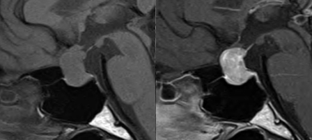
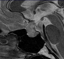
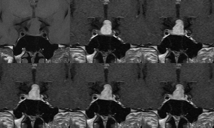
| Discussion | Pituicytoma, granular cell tumor of the sellar region, and spindle cell oncocytoma (follow the WHO CNS5) Imaging findings There is a well-defined mass involving sellar and suprasellar region. The mass is isointense with brain on T1WI and hyperintense on T2WI. On dynamic contrast enhanced studies, it is clearly separate from anterior pituitary gland, which shows delayed enhancement. It shows fast and relatively homogeneous enhancement. Pituitary stalk is not well delineated. 고찰 Pituicytoma, granular cell tumor of sellar region, spindle cell oncocytoma are included in pituicyte tumor family. They arise from modified glial cells (pituicytes) that reside in the infundibular stalk and neurohypophysis. Radiographic features : CT - homogeneously enhancing mass - either within pituitary fossa or in the suprasellar region : MRI - intrasellar mass that is clearly separate from anterior pituitary gland and enhances homogeneously is most likely a pituicytoma - T1: isointense solid mass - T2: hyperintense - T1 C+ (Gd): homogeneous contrast enhancement |
| Correct answer |
Correct Answer
Semi-Correct Answer |