HOME > Weekly Case > Weekly Case
Weekly Case
종결
Case No. 604
2021-10-01~2021-10-31
출제자 : 류경화
Hit : 593
| Period | 2021-10-01~2021-10-31 |
|---|---|
| Diagnosis | Solitary fibrous tumor |
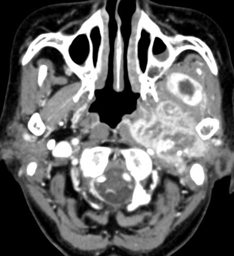
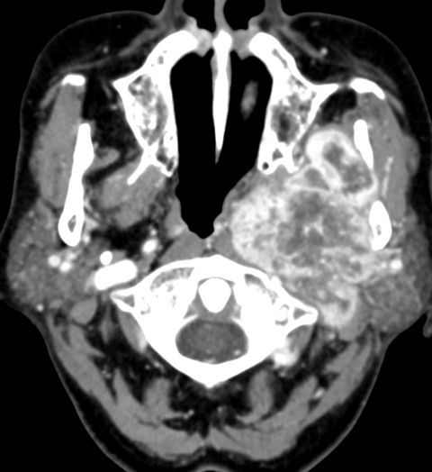
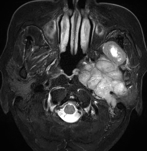
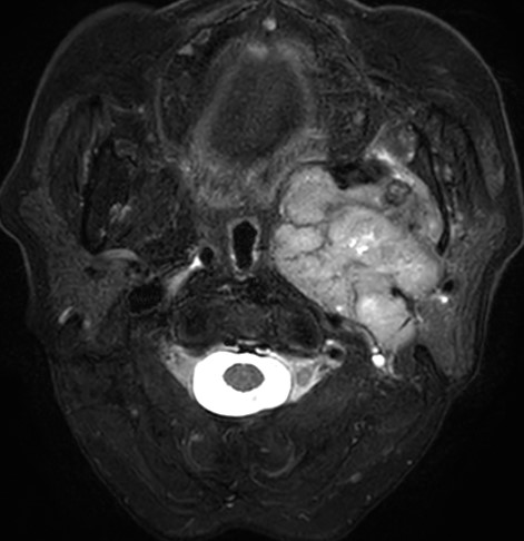
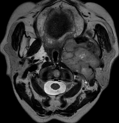
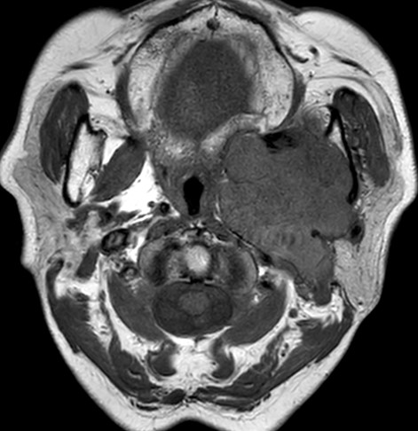
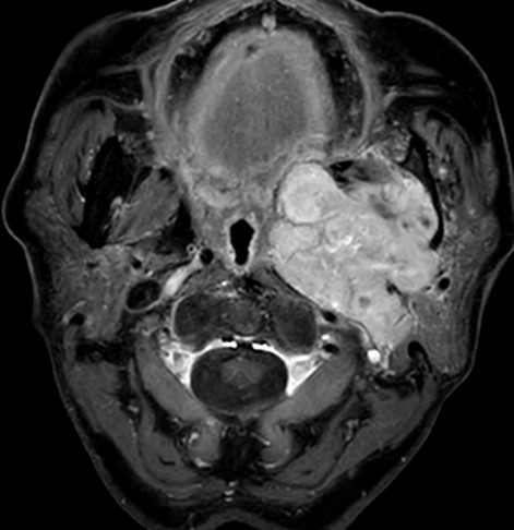
| Clinical information | 79Y/F Brain MRI에서 우연히 발견되어 work up 하게 된 병변임. 진단은? |
| Discussion | Solitary fibrous tumor of parapharyngeal space • Rare, but well-known mesenchymal origin tumor • Composed mainly of relatively uniform spindle cells in collagenous stroma • Location -Pleura -Other than the pleura: retroperitoneum, deep soft tissues of the extremities, abdominal cavity, head and neck (infratemporal fossa, parapharyngeal space, nasal and paranasal sinuses, soft palate, and epiglottis) • Symptom of parapharyngeal SFT: minimal, bulge in the overlying pharynx • CT finding -Round, well-defined and isolated mass -Calcifications • MRI finding -Well-defined, lobulated mass -Isointense on T1WI, hyperintense on T2WI, prominent and heterogeneous enhancement -Cystic and myxoid degeneration : Focal areas of T1 low, T2 high signal -Hypervascularity of the tumor: Heterogeneous and intense contrast enhancement in both CT and MR |
| Correct answer |
Correct AnswerSemi-Correct Answer |