HOME > Weekly Case > Weekly Case
Weekly Case
종결
Case No. 602
2021-09-01~2021-09-30
출제자 : 박미나
Hit : 732
| Period | 2021-09-01~2021-09-30 |
|---|---|
| Diagnosis | Cavernous Sinus Hemangioma |
| Gender |
|
| Age | 58 |
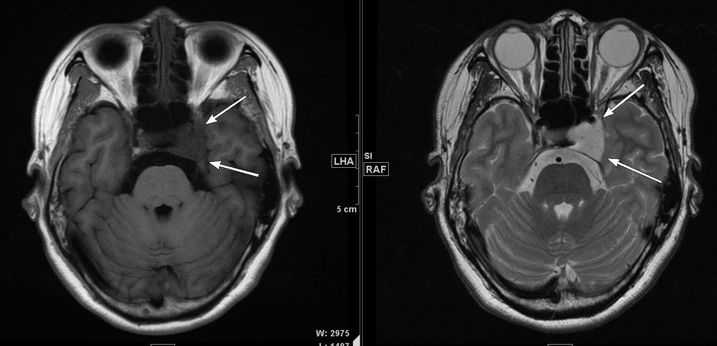
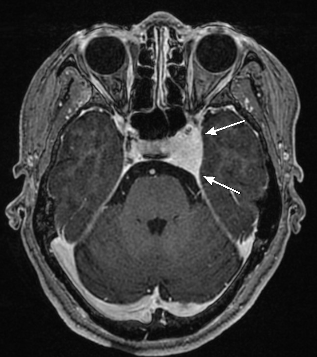
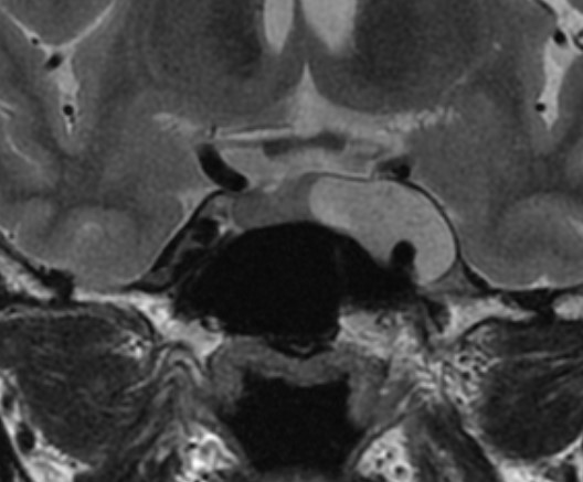
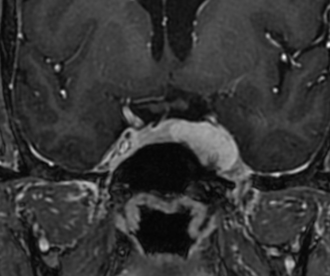
| Clinical information | 58세 여자로 MRI 상 우연히 발견된 병변이다. 진단은? |
| Discussion | ** Cavernous Sinus Hemangioma - uncommon benign lesions. - fewer than 1% of parasellar masses, with a ratio F:M = 7:1. - usually seen in the fourth decade of life Sx: headaches and retro-orbital pain to cranial nerve palsies with ophthalmoplegia. Imaging findings: - a well-defined mass with low to isointensity on T1-weighted images - marked hyperintense signal on T2-weighted images - progressive enhancement with a centripetal fill-in pattern. Diagnosis: a labeled red cell blood pool scintigraphy. |
| Correct answer |
Correct Answer
Semi-Correct Answer |