HOME > Weekly Case > Weekly Case
Weekly Case
종결
Case No. 596
2021-07-01~2021-07-31
출제자 : 박예원
Hit : 705
| Period | 2021-07-01~2021-07-31 |
|---|---|
| Diagnosis | MVNT (multinodular vaculoated neuronal tumor) |
| Gender |
|
| Age | 44 |
| Clinical information | F/44 CC: incidental lesion (검진) |
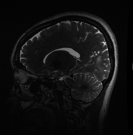
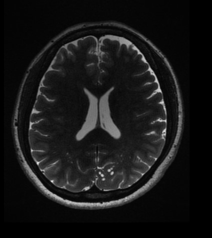
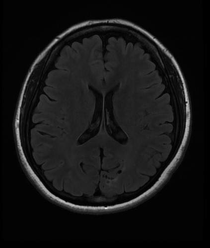
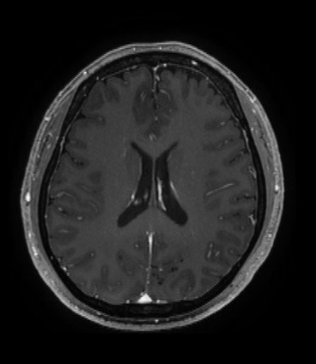
| Discussion | 1) Imaging finding Multple clusters of T2 high nodular lesions at the left parietooccipital lobe subcortex, with surrounding white matter hyperintensity. - no enhancement. -> Most likely MVNT. 2) Facts Multinodular and vacuolating neuronal tumor of the cerebrum is a benign, mixed glial neuronal lesion that is included in the 2016 updated World Health Organization classification of brain neoplasms as a unique cytoarchitectural pattern of gangliocytoma. MVNTs exhibit a highly characteristic appearance on MR imaging. The manifest as a cluster of variably sized nodular lesions located on the subcortical ribbon and superficial subcortical white matter following the gyral contour. They appear hyperintense on T2WI, do not suppress on FLAIR, and typically do not enhance or cause mass effect. While a few MVNTs may be causative of seizure disorders and therefore require excision for clinical management, akin to DNET, most are asymptomatic lesions that are “leave me alone” entities. These lesions usually do not require biopsy, resection, or follow-up imaging unless and until it can be demonstrated that the visualized lesion is responsible for the symptoms. |
| Correct answer |
Correct Answer
Semi-Correct Answer |