HOME > Weekly Case > Weekly Case
Weekly Case
종결
Case No. 595
2021-07-01~2021-07-31
출제자 : 박예원
Hit : 530
| Period | 2021-07-01~2021-07-31 |
|---|---|
| Diagnosis | Pilocytic astrocytoma |
| Gender |
|
| Age | 21 |
| Clinical information | M/21 CC: left side weakness 진단은? |
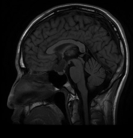
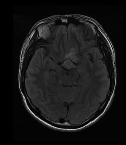
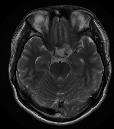
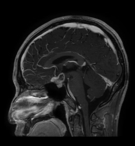
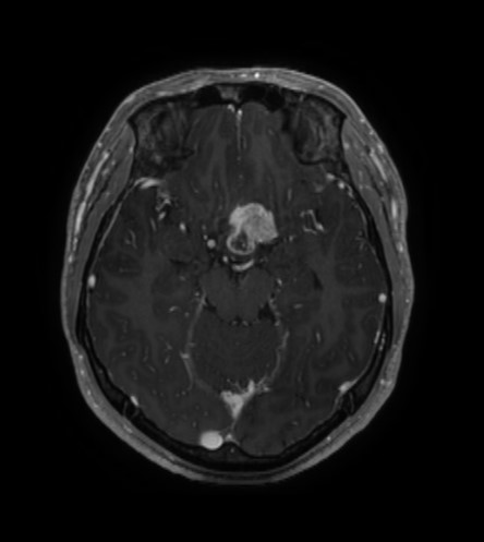
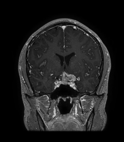
| Discussion | 1) Imaging finding: An approx 2.4-cm mass involving suprasellar cisern, extending to the 3rd ventricular floor and tuberculum sella - partially encasing the left supraclinoid ICA, proximal M1 and A1 without stenosis. - posteriorly compressing the optic chiasm. - pituitary stalk is not well delineated. - DDx> suprasellar mass such as craniopharyngioma, tuberculum sella meningioma, pilocytic astrocytoma, etc.. 2) facts Pilocytic astrocytoma (PA) is classified as World Health Organization (WHO) grade I tumor, which occurs most commonly in the pediatric population. However, it also accounts for approximately 0.8-1.5% of adult brain tumors, and up to 25% of patients with PA are older than 20 years. Identifying pediatric PAs is relatively simple, because majority of tumors show typical cerebellar location with ‘cystic mass with mural nodule appearance’. On the other hand, identifying adult PAs is confusing due to the fact that approximately half of adult PAs show a supratentorial location, and that they manifest large spectrum of imaging presentations, which often show aggressive imaging features. It should be kept in mind that PAs can also show location at the optic nerve and chiasm, hypothalamus, and in this location the ‘cystic mass with mural nodule appearance’ is rarely manifestated. Although it is difficult, sometimes almost impossible to DDx the lesion with other common suprasellar lesions such as craniopharyngioma or meningioma, the diagnosis should be kept in mind. |
| Correct answer |
Correct Answer
Semi-Correct Answer |