HOME > Weekly Case > Weekly Case
Weekly Case
종결
Case No. 594
2021-07-01~2021-07-31
출제자 : 박예원
Hit : 411
| Period | 2021-07-01~2021-07-31 |
|---|---|
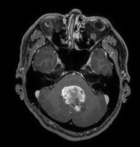
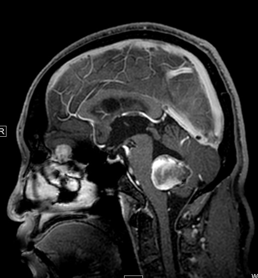
| Diagnosis | partially thrombosed intraventricular PICA aneurysm |
| Gender |
|
| Age | 54 |
| Clinical information | M/54 CC: posterior neck pain (for 2.5 years) 가능한 진단은? |
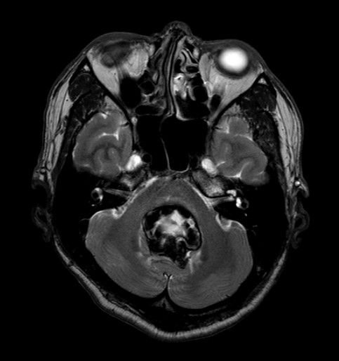
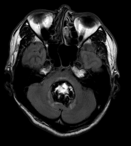
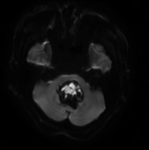
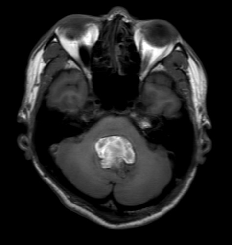
| Discussion | 1) Imaging finding: An approx 3.6-cm pre T1 high lesion with focal small enhancing portion at the 4th ventricle. - with hydrocephalus and perilesional edema. - internal hemorrhage Note: retrospective MRI review상 PICA와의 연결성을 정확히 tracing하기는 어려웠음. Op. finding상 intraventricular partially thrombosed giant aneurysm이 관찰되었으며, clipping 시행함. 2) facts Distal PICA aneurysms are uncommon and their pathogenesis is a matter of debate. While most intracranial aneurysms are located at arterial bifurcations, distal PICA lesions arise predominantly from the arterial side wall of the telovelotonsillary segment. It was proposed that increased hemodynamic wall shear stress induced by the hairpin curvature of the cranial loop within this segment is responsible for aneurysm formation. Our patient demonstrates how a large partially thrombosed distal PICA aneurysm can mimic an infratentorial tumor both clinically and radiologically. Most large distal PICA aneurysms rarely rupture and seldom cause thromboembolic events due to the rich anastomotic arterial supply of the inferior cerebellum. Their presentation is similar to posterior fossa neoplasms inducing chronic brainstem or cerebellar compression. Giant partially thrombosed/thrombosed aneurysms arising from the distal PICA are rare, but should be kept in mind as a differential diagnosis of 4th ventricular lesions. |
| Correct answer |
Correct Answer
Semi-Correct Answer |