HOME > Weekly Case > Weekly Case
Weekly Case
종결
Case No. 589
2021-05-01~2021-05-31
출제자 : 이아름
Hit : 787
| Period | 2021-05-01~2021-05-31 |
|---|---|
| Diagnosis | Uremic encephalopathy |
| Gender |
|
| Age | 57 |
| Clinical information | 57/M C.C: DM, ESRD, mental change 가장 가능성이 높은 진단은? |
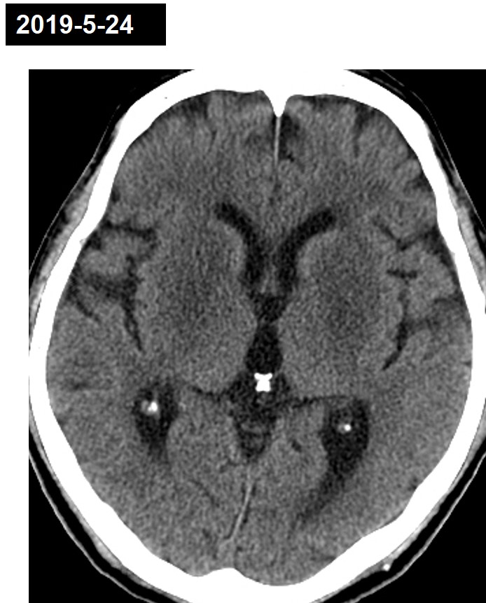
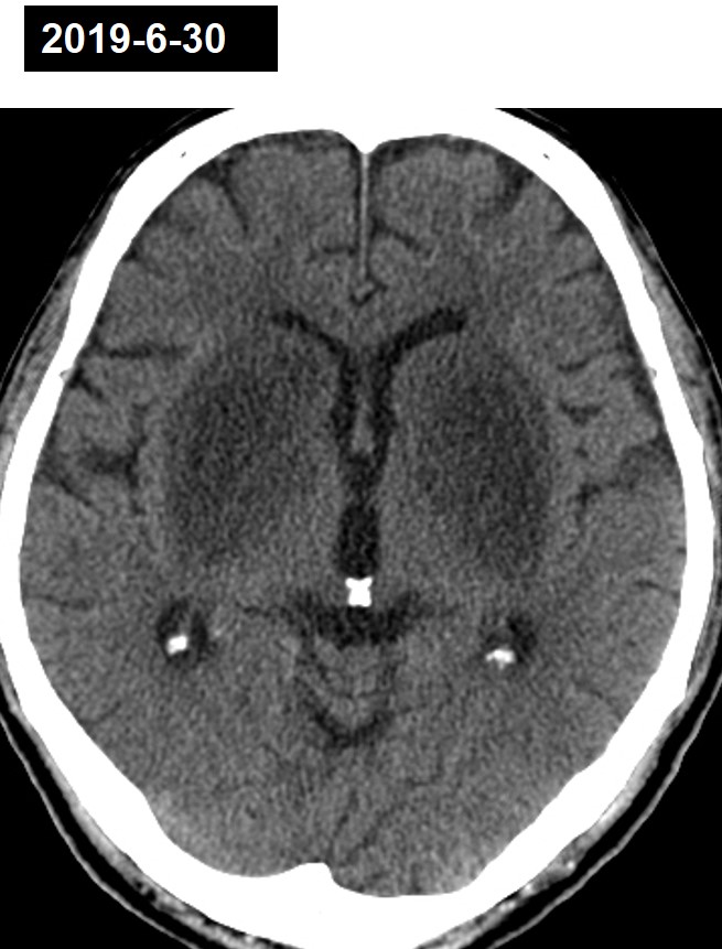
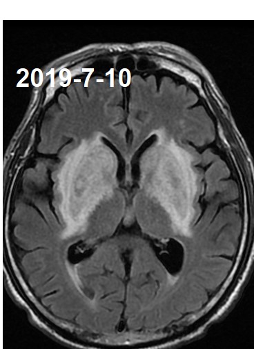
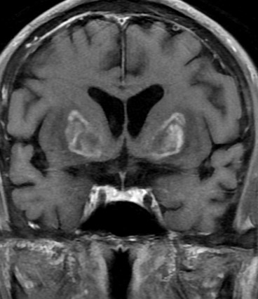
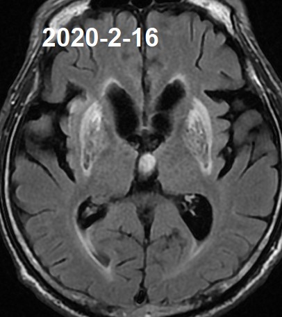
| Discussion | Imaging finding - CT: More progressive symmetric enlargement of low density in the bilateral basal ganglia with swelling. --> Probable aggravation of uremic encephalopathy in DM patient. - MRI: Again noted bilateral symmetrical T2 high intensity lesions in the bilateral BG including globus pallidus, putamen and head of caudate nucleus, external capsule and internal capsule. On follow up MRI, atrophic change with diffuse T2 high SI and mild cerebromalacic change in bilateral BG including globus pallidus, putamen and head of the caudate nucleus, external capsule and internal capsule, since previous MRI -- Chronic change with atrophy due to uremic encephalopathy associated with ESRD state. Uremic encephalopathy 1) Alterations of the bilateral basal ganglia in the acute stage - Basal ganglia are particularly vulnerable to a wide range of toxins and metabolic changes - Worsening of the renal condition cause of the acute exposure of the basal ganglia to uremic toxins 2) By longstanding DM - cerebrovascular reactivity impairment - endothelial dysfunction in cerebral vessels 3) Hyperglycemia by itself - Impairment of the endothelium-dependent vasoreactivity of cerebral arterioles - Focal destructive endothelial lesions, contributes to blood–brain barrier breakdown - Increases free radical release |
| Correct answer |
Correct Answer
Semi-Correct Answer |