HOME > Weekly Case > Weekly Case
Weekly Case
종결
Case No. 586
2021-04-01~2021-04-30
출제자 : 정미선
Hit : 552
| Period | 2021-04-01~2021-04-30 |
|---|---|
| Diagnosis | Progressive multiple leukoencephalopathy |
| Gender |
|
| Age | 40 |
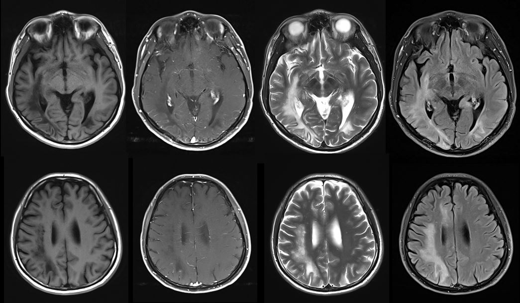
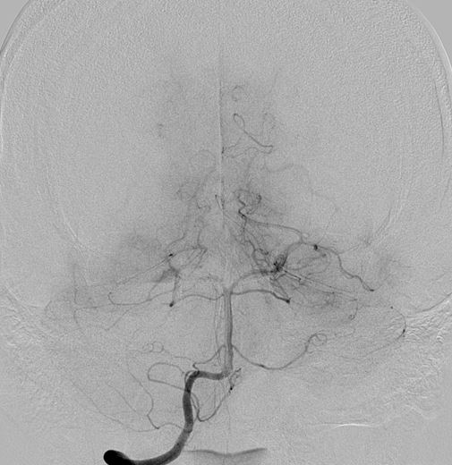
| Clinical information | 40/M 왼쪽 시야장애, 어지러움 Lab: WBC, 2500/μl 진단은? |
| Discussion | Progressive multiple leukoencephalopathy - A demyelinating disease which results from the reactivation of John Cunningham virus (JC virus) infecting oligodendrocytes in patients with compromised immune systems. - PML occurred in patients with AIDS, typically developing in patients with CD4 counts of 50-100 cells/μL, and is found in approximately 5% of autopsies of patients who died from AIDS # Image findings - CT: Asymmetric low attenuation involving the periventricular and subcortical white matter. - MRI : Multifocal, asymmetric periventricular and subcortical T2/FLAIR high SI, T1 hypointense lesions without enhancement, the subcortical U-fibers involvement with a predilection for the parieto-occipital area. : Peripheral patchy diffusion restriction particularly at the leading edge. : Elevated perfusion at the leading edge of lesions (most commonly seen in progressive non-IRIS PML) |
| Correct answer |
Correct Answer
Semi-Correct Answer |