HOME > Weekly Case > Weekly Case
Weekly Case
종결
Case No. 579
2021-02-01~2021-02-28
출제자 : 배윤정
Hit : 345
| Period | 2021-02-01~2021-02-28 |
|---|---|
| Diagnosis | Bing Neel syndrome (BNS) |
| Gender |
|
| Age | 70 |
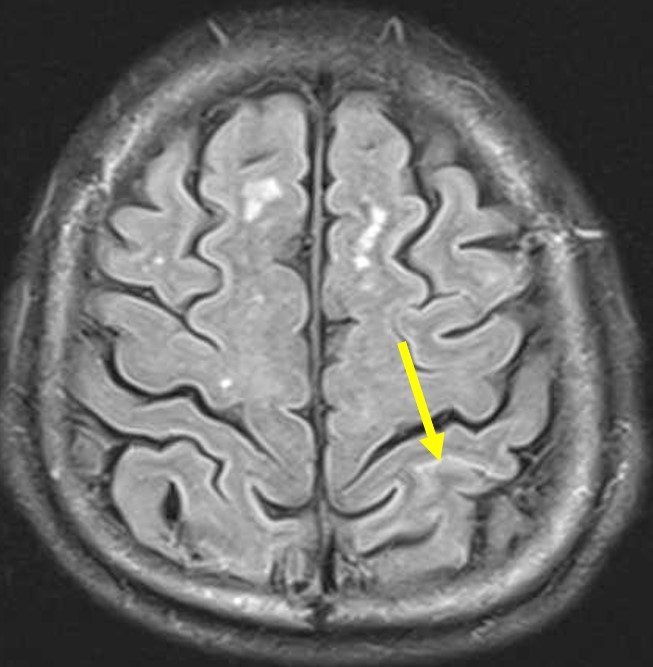
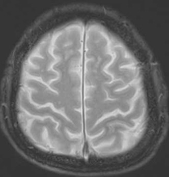
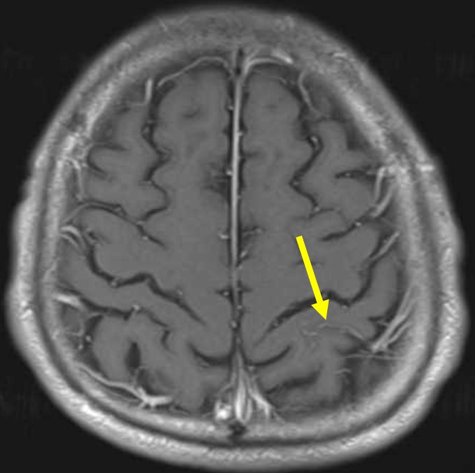
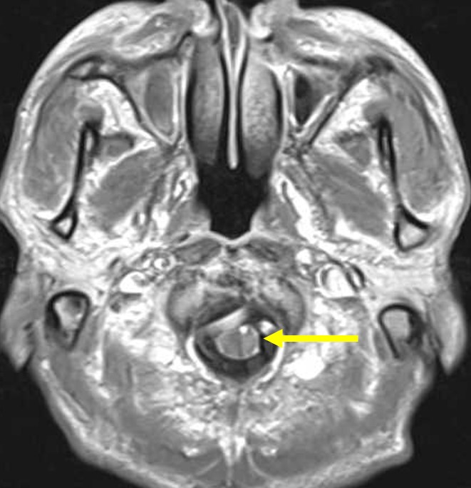
| Clinical information | M/70Y Symptom: Headache History: Diagnosed with Waldenström’s macroglobulinemia What is your diagnosis? |
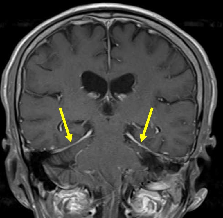
| Discussion | Bing Neel syndrome (BNS) Definitions: - A rare disease manifestation of Waldenström’s macroglobulinemia (WM) - Malignant lymphoplasmacytic cells invade the central nervous system (CNS), and may be detected in the cerebrospinal fluid, the meninges, and/or the cerebral parenchyma. Clinical presentations: - Symptoms are diverse and reflect involvement of the CNS including headache, nausea and vomiting, visual disturbances, hearing loss and cranial neuropathies, seizures, cognitive decline, aphasia, psychiatric symptoms, cerebellar dysfunction, impairment of consciousness including coma, and paresis typically represent involvement of brain parenchyma or the spinal cord. Diagnosis: - The golden standard for the diagnosis of BNS is a histological biopsy of the cerebrum or meninges - When there is leptomeningeal involvement, the CSF may contain malignant lymphoplasmacytic cells. It is therefore recommended to perform repeated CSF analysis and, if possible, to do so before MRI is performed to exclude non-specific meningeal enhancement that occurs after CSF sampling. MRI findings: - MRI of the brain and spinal cord is advised in cases of suspected BNS. The goal of neuroimaging is not only to find supportive evidence for BNS, but also for the exclusion of differential diagnoses (infectious and others), and to select a possible site for biopsy. -There are two forms of CNS involvement by BNS: 1. The diffuse form. -- Lymphoid cell infiltration in the leptomeningeal sheaths and the perivascular spaces -- Presents with contrast enhancement and/or thickening of meningeal sheaths 2. The tumoral form -- Can be unifocal or multifocal, and is usually located in the deep subcortical hemispheric regions -- Increased parenchymal signal intensity can be identified in T2 and in FLAIR images, and corresponding contrast-enhancement of the lesion can be seen. Treatment options: - Steroid, chemotherapy, Anti-CD20 therapy - Contrast-enhancing lesions may respond well to the treatment. |
| Correct answer |
Correct Answer
Semi-Correct Answer |