HOME > Weekly Case > Weekly Case
Weekly Case
종결
Case No. 576
2021-01-01~2021-01-31
출제자 : 서종현
Hit : 530
| Period | 2021-01-01~2021-01-31 |
|---|---|
| Diagnosis | Anti-MOG antibody encephalitis |
| Gender |
|
| Age | 73 |
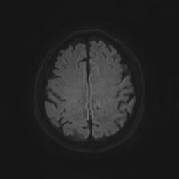
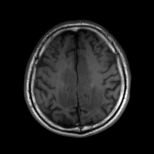
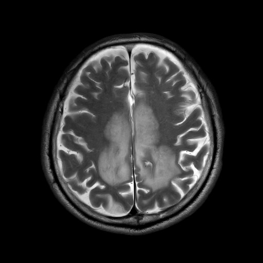
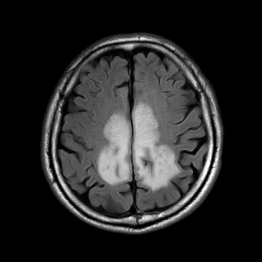
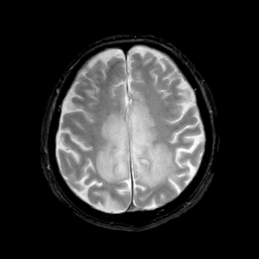
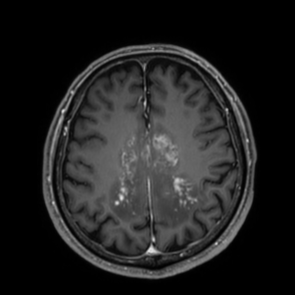
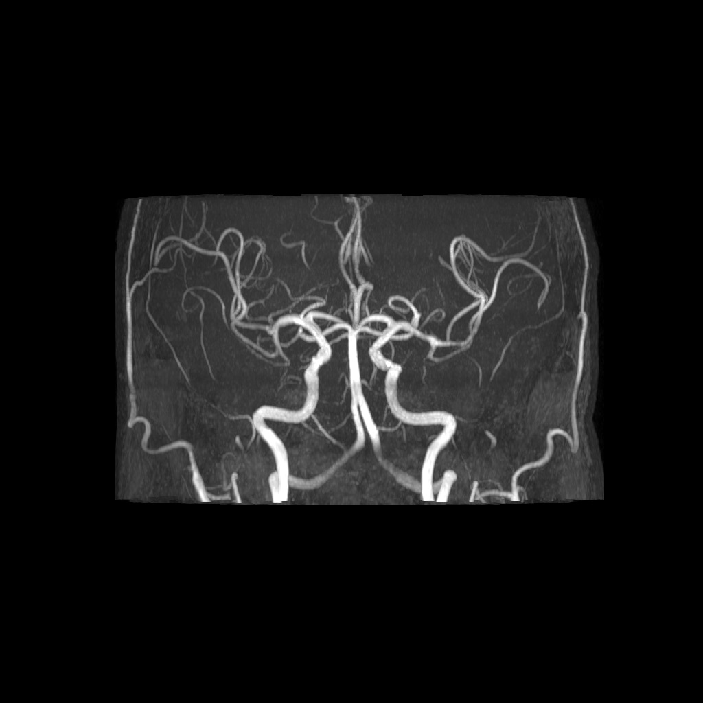
| Clinical information | 10일전부터 발생한 ataxia를 주소로 내원한 73세 남자환자이다. DM, HTN, Heart failure있고, 다른 기저질환 없는 환자이다. 진단은? |
| Discussion | Anti-MOG antibody encephalitis Brain stereotactic biopsy상 perivascular lymphocytic aggregation로 나왔고 anti-MOG Ab 양성 나옴. 특별한 치료 하지 않고 1달반 후 f/u에서 호전됨. 1. Introduction Myelin oligodendrocyte glycoprotein antibody (MOG-Ab) associated disease - New spectrum of autoimmune disorders with Abs against the MOG predominantly involving the optic nerve and spinal cord - MOG: a glycoprotein located on the myelin surface 2. Clinical manifestation Optic neuritis (54-61%), myelitis (44-83%), encephalitis Pathologic findings can mimic vasculitis. - Presence of lymphoid cells in vessel walls must be interpreted with caution. 3. Radiologic Findings: Brain Normal in 2/3 of cases Bilateral lesions at onset, fee (<4) lesions Nonspecific T2 high SI foci in supratentorial subcortical or deep WM Rhombencephalitis: subtentorial lesion (1/3) ADEM-like diffuse signal changes in the cortical GM, subcortical/deep WM and deep GM on T2WI and FLAIR - Poorly delineated, subtle multiple patch enhance, “cloud-like” - Scattered linear and nodular enhancement (active lesions) with some restricted diffusion Leptomeningeal enhancement and thalamic lesions were unique. Tumefactive lesion: poorly demarcated lesions and cortical lesions |
| Correct answer |
Correct AnswerSemi-Correct Answer |