HOME > Weekly Case > Weekly Case
Weekly Case
종결
Case No. 572
2020-11-01~2020-11-30
출제자 : 김슬기
Hit : 617
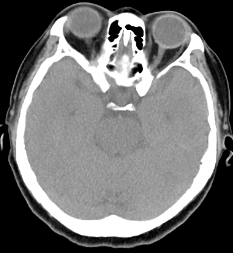
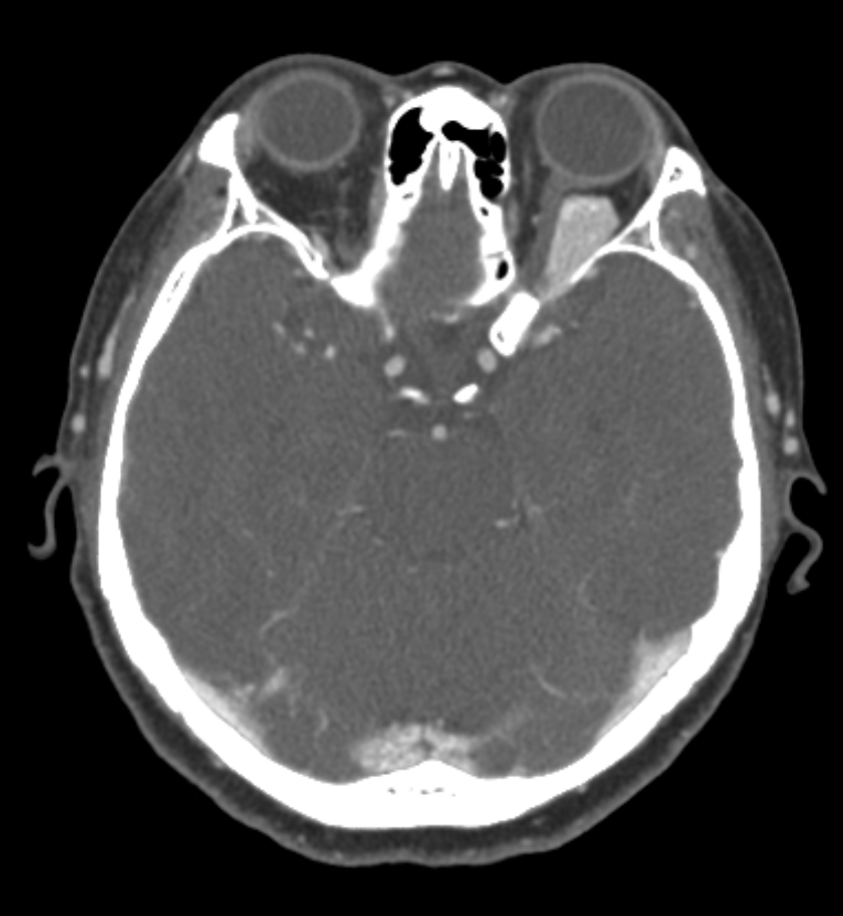
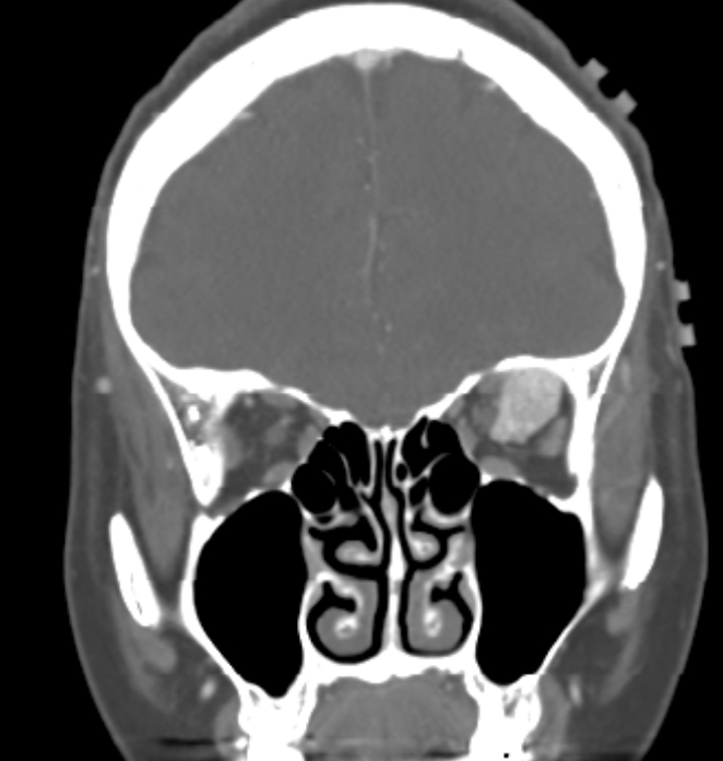
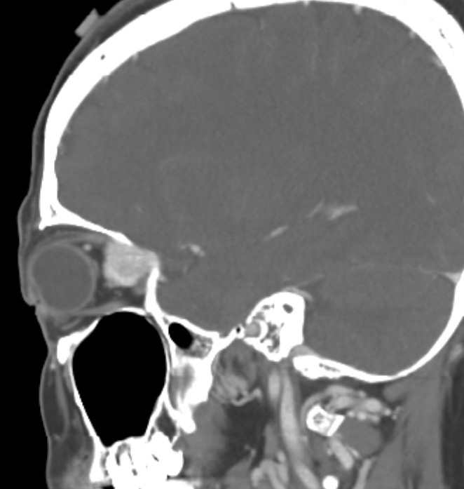
| Period | 2020-11-01~2020-11-30 |
|---|---|
| Diagnosis | Orbital venous varix |
| Gender |
|
| Age | 50 |
| Clinical information | 50세 여자 Left ocular pain을 주소로 내원. 진단은? |
| Discussion | Radiographic features : Can be difficult or impossible to detect without Valsalva maneuver. Contrast-enhanced studies will demonstrate venous phase opacification. CT - If the diagnosis is suspected, CT should be performed both at rest and with a Valsalva maneuver - Irregular or smooth enhancing lesion which significantly increases in size with straining, typically located at the orbital apex - Enhancement match that of other venous structures - In the setting of thrombosis, the enhancement may be absent, and no change on the Valsalva maneuver MRI In the absence of thrombosis: T1 & T2: hypointense c.f. extraocular muscles If thrombosis is present the imaging is more variable T1 & T2: heterogeneous with areas of high signal |
| Correct answer |
Correct Answer
Semi-Correct Answer |