HOME > Weekly Case > Weekly Case
Weekly Case
종결
Case No. 531
2019-10-01~2019-10-31
출제자 : 김슬기
Hit : 274
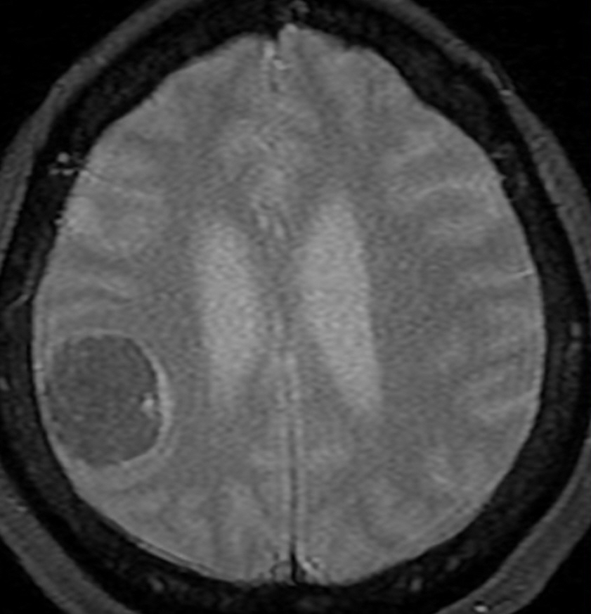
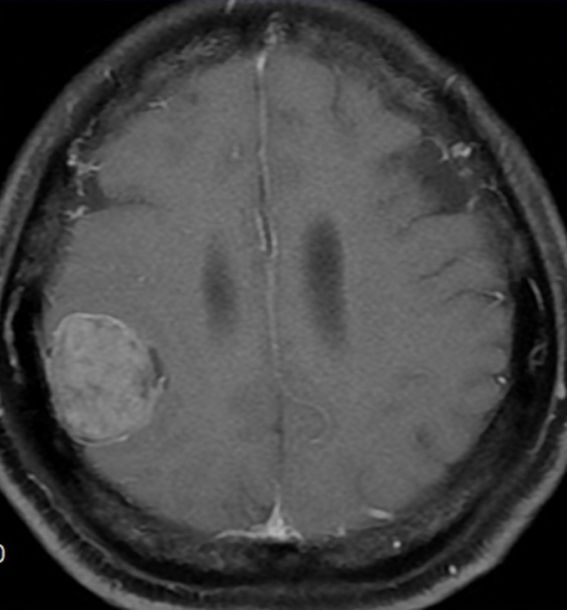
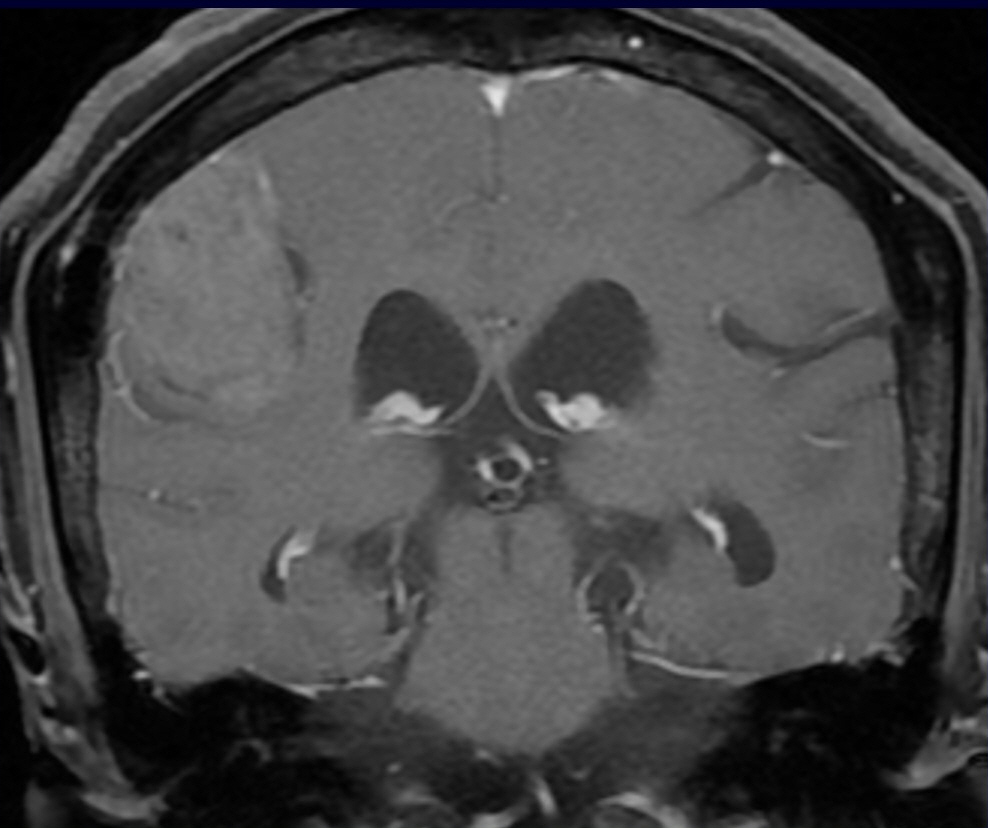
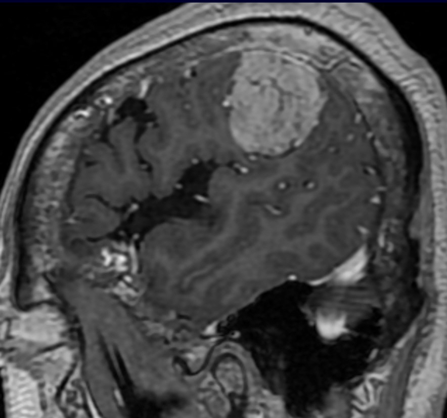
| Period | 2019-10-01~2019-10-31 |
|---|---|
| Diagnosis | Solitary fibrous tumor/Hemangiopericytoma |
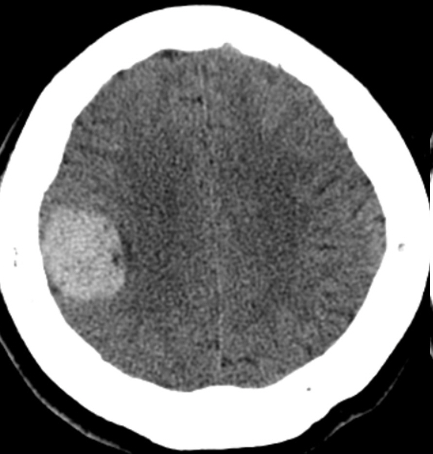
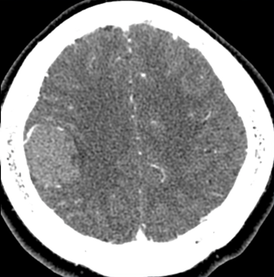
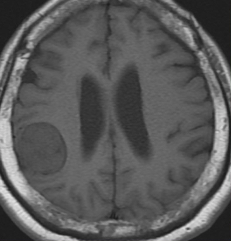
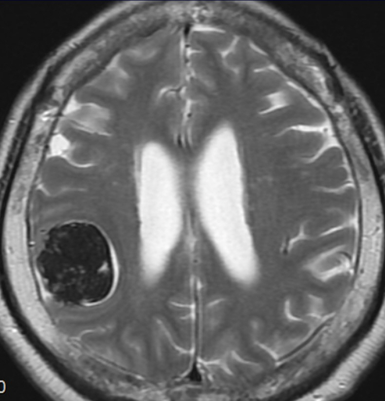
| Clinical information | 56세 여자 우연히 발견된 뇌의 종괴를 주소로 내원함. 유방암 수술 과거력 있음 진단은? |
| Discussion | **Solitary fibrous tumor/Hemangiopericytoma -Rare, but most common CNS nonmeningothelial mesenchymal tumor -SFT and HPC share the same molecular genetic profile -> 2016 WHO has created the combined term solitary fibrous tumor/hemangiopericytoma (SFT/HPC) -Highly cellular and vascularized mesenchymal tumor, nearly always attached to dura / Extra-axial tumor -SFTs/HPCs are almost always solitary, usually supratentorial masses, often lobulated in contour. They are highly vascular and have a tendency to erode adjacent bone -Imaging features - SFT/HPC grade I hypointense on T2WI - SFT/HPC II resembles aggressive meningioma • Calcification absent • Iso- to mildly hyperintense on T1WI • Iso- to heterogeneously hyperintense on T2WI • Very vascular, prominent "flow voids" • Enhances strongly but heterogeneously • "Dural tail" sign usually absent |
| Correct answer |
Correct AnswerSemi-Correct Answer |