HOME > Weekly Case > Weekly Case
Weekly Case
종결
Case No. 529
2019-09-01~2019-09-30
출제자 : 박미나
Hit : 473
| Period | 2019-09-01~2019-09-30 |
|---|---|
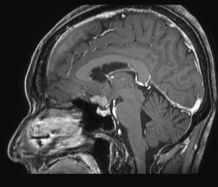
| Diagnosis | Intracranial gossypiboma |
| Gender |
|
| Age | 59 |
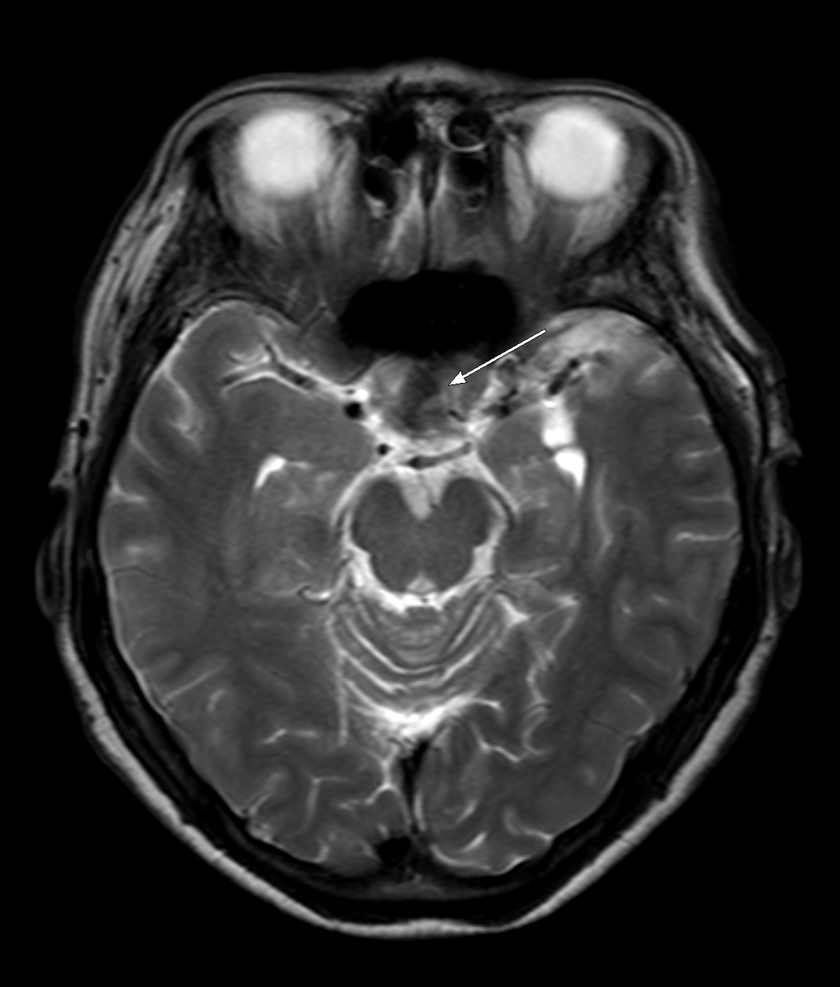
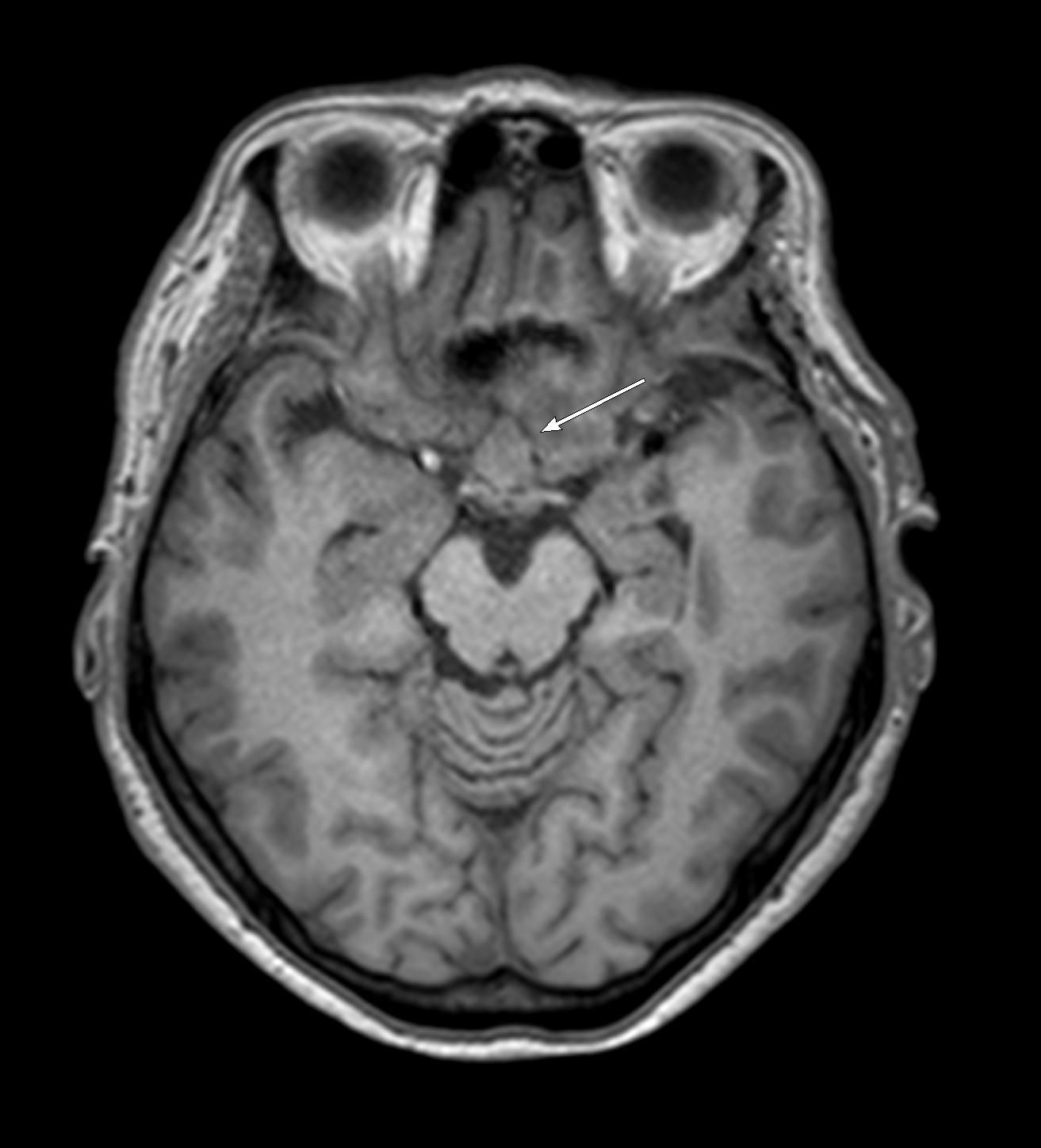
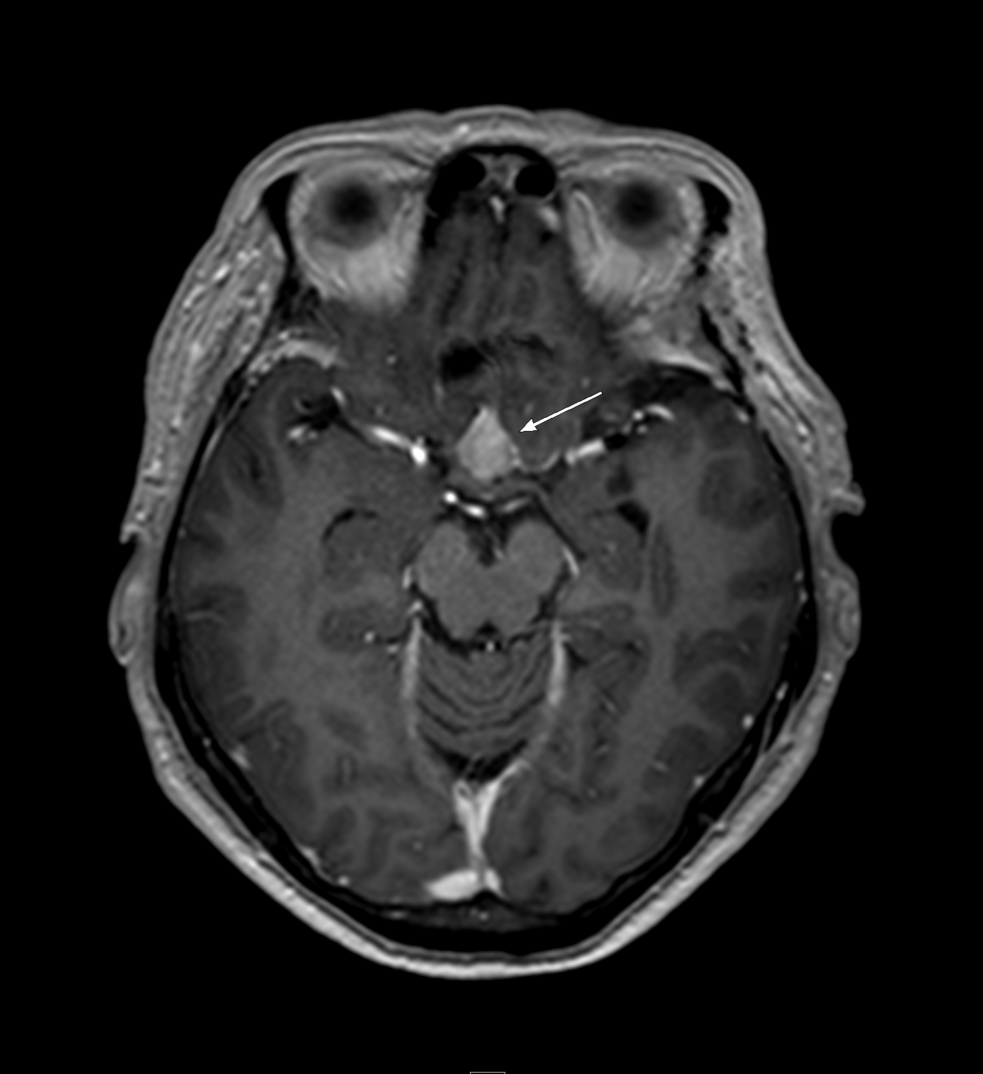
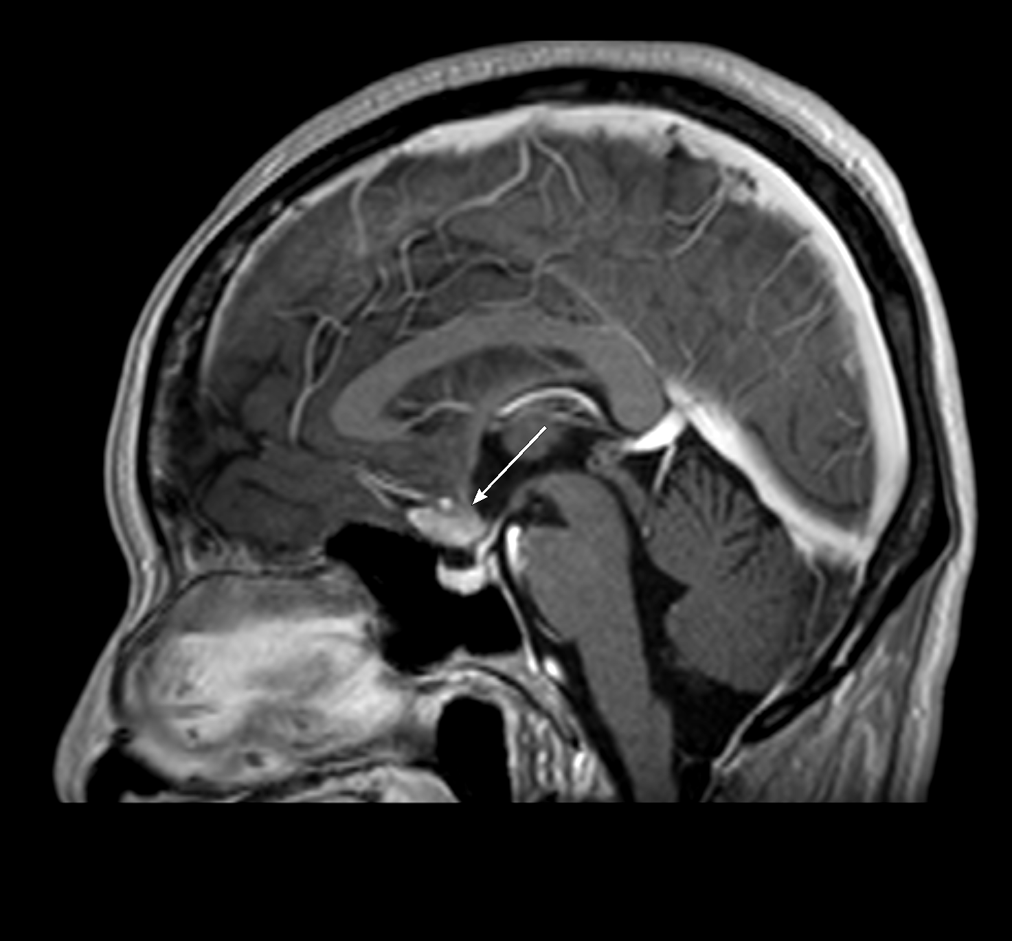
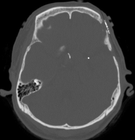
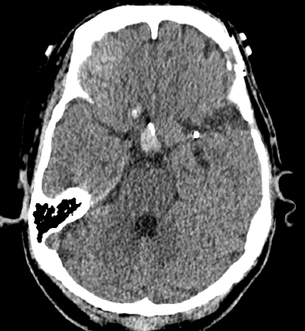
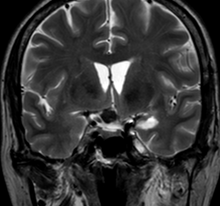
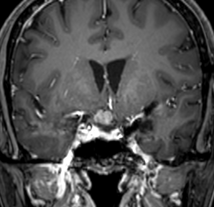
| Clinical information | 59세 여환 Hx> 외부병원에서 6개월전 tuberculum sellae meningioma 로 수술 후 추적 검사로 시행한 MR과 3개월 추적 검사 MR 이다. 진단은? |
| Discussion | Intracranial gossypiboma Gossypiboma is a term used to describe a foreign-body reaction surrounding material made of cotton matrix. Imaging is essential to the workup for gossypibomas, and the common differential diagnosis includes primary, metastatic, recurrent tumor, radiation necrosis or abscess. Suggested CT findings are low-density heterogeneous masses with a hyperdense rim that was further enhanced after contrast injection, and sometimes they present with a spongiform pattern containing air bubbles. The signal intensity of MRI may vary according to aging, histologic composition, and its fluid content and gossypibomas cannot be easily distinguished from a recurrent tumor or abscess due to their less characteristics. |
| Correct answer |
Correct Answer
Semi-Correct Answer |