HOME > Weekly Case > Weekly Case
Weekly Case
종결
Case No. 525
2019-08-01~2019-08-31
출제자 : 강경미
Hit : 293
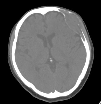
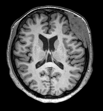
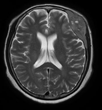
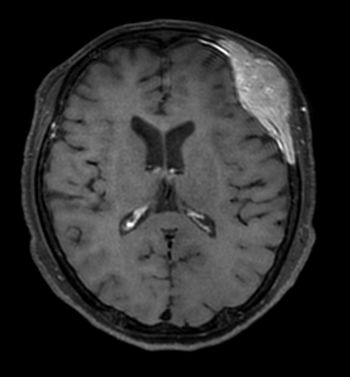

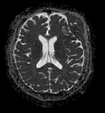
| Period | 2019-08-01~2019-08-31 |
|---|---|
| Diagnosis | Intraosseous meningioma |
| Gender |
|
| Age | 73 |
| Clinical information | F/73 Left frontal scalp swelling/tenderness(6MA) 진단은? |
| Discussion | • Rare subtype of meningioma, most common type of extradural meningioma • Benign slowly growing tumor • Common site : Frontoparietal, orbital regions • Imaging Findings – CT • osteoblastic (60%), osteolytic (30%), or mixed type. • hyperostosis, atypical vascular marking, irregular foci of calcification – MRI • T1: isointense extra-axial mass component with the expanded bony component being low signal similar to the rest of the skull • T2: isointense to grey matter while a small proportion can be hyperintense • T1 C+ (Gd): uniform avid contrast enhancement, dural enhancement. • Differential diagnosis – Craniofacial fibrous dysplasia • Young age, stops growing after puberty • Inner table of the skull looks smooth – Osteoma: non-enhancing, dense, smooth, and well-demarcated lesions. – Osteosarcoma, Paget disease, Osteoblastic metastasis • Treatment – Wide surgical resection with cranial reconstruction – Non-resectable : gamma knife, chemotherapy, and disphosphonate therapy |
| Correct answer |
Correct Answer
Semi-Correct Answer |