HOME > Weekly Case > Weekly Case
Weekly Case
종결
Case No. 507
2019-02-01~2019-02-28
출제자 : 배윤정
Hit : 549

| Period | 2019-02-01~2019-02-28 |
|---|---|
| Diagnosis | ARIA-E (Amyloid Related Imaging Abnormalities) |
| Gender |
|
| Age | 69 |
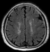
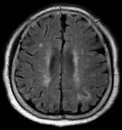
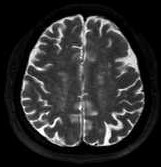
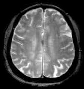
| Clinical information | F/69Y Under medication for Alzheimer’s disease Patient’s history: -On amyloid-modifying therapeutic agents Fig1 - initial FLAIR, Fig2-5 - follow up MRI What is your diagnosis on follow-up MRI? |
| Discussion | ARIA-E (Amyloid Related Imaging Abnormalities) MRI: -On FLAIR image, increased MR signal in left parietal sulcus -Absence of hemorrhage Patient’s history: -On amyloid-modifying therapeutic agents ARIA (Amyloid Related Imaging Abnormalities) -ARIA is the spectrum of MRI abnormalities including signal changes thought to represent vasogenic edema and microhemorrhage, which is related to amyloid-modifying therapies. -ARIA-E: a. ARIA-E refers to the MR signal alterations from vasogenic edema and extravasated fluid phenomena. b. Increased MR signal intensity on FLAIR or other T2-weighted sequences in the parenchyma and/or leptomeninges in the parietal, occipital, and frontal lobes, but also can be observed in the cerebellum and brainstem. c. In some cases, the increased MR signal can be seen in sulcal space due to effusion or proteinaceous collection of fluid. d. This can be misinterpreted as subarachnoid hemorrhage, but no evidence of blood product. -ARIA-H: a. ARIA-H refers to the MR signal alterations on attributable to microhemorrhage and hemosiderosis. b. Microhemorrhage and/or hemosiderosis can be detected on long echo time gradient refocused echo sequences (T2*-GRE) or susceptibility weighting imaging (SWI). c. This is thought to represent residua of a small leakage of blood from a vessel into adjacent tissue parenchyma and deposition of iron. |
| Correct answer |
Correct Answer
Semi-Correct Answer |